

Automatic Detection of the Inner Ears in Head CT Images Using Deep Convolutional Neural Networks
- PMID: 31007337
- PMCID: PMC6474381
- DOI: 10.1117/12.2293383
Automatic Detection of the Inner Ears in Head CT Images Using Deep Convolutional Neural Networks
Abstract
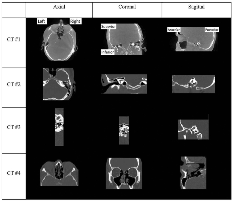
Cochlear implants (CIs) use electrode arrays that are surgically inserted into the cochlea to stimulate nerve endings to replace the natural electro-mechanical transduction mechanism and restore hearing for patients with profound hearing loss. Post-operatively, the CI needs to be programmed. Traditionally, this is done by an audiologist who is blind to the positions of the electrodes relative to the cochlea and relies on the patient's subjective response to stimuli. This is a trial-and-error process that can be frustratingly long (dozens of programming sessions are not unusual). To assist audiologists, we have proposed what we call IGCIP for image-guided cochlear implant programming. In IGCIP, we use image processing algorithms to segment the intra-cochlear anatomy in pre-operative CT images and to localize the electrode arrays in post-operative CTs. We have shown that programming strategies informed by image-derived information significantly improve hearing outcomes for both adults and pediatric populations. We are now aiming at deploying these techniques clinically, which requires full automation. One challenge we face is the lack of standard image acquisition protocols. The content of the image volumes we need to process thus varies greatly and visual inspection and labelling is currently required to initialize processing pipelines. In this work we propose a deep learning-based approach to automatically detect if a head CT volume contains two ears, one ear, or no ear. Our approach has been tested on a data set that contains over 2,000 CT volumes from 153 patients and we achieve an overall 95.97% classification accuracy.
Keywords: Cochlear implant; Convolutional Neural Networks; image classification; image-guided cochlear implant programming.
Figures


References
-
- NIDCD Fact Sheet: Cochlear Implants, “National institute on deafness and other communication disorders,” NIH Publication No. 11–4798, https://www.nidcd.nih.gov/sites/default/files/Documents/health/hearing/F... (2011).
Grants and funding
LinkOut - more resources
Full Text Sources
Research Materials
Miscellaneous