Applicability of Trichoscopy in Scalp Seborrheic Dermatitis
- PMID: 31007472
- PMCID: PMC6463453
- DOI: 10.4103/ijt.ijt_86_18
Applicability of Trichoscopy in Scalp Seborrheic Dermatitis
Abstract
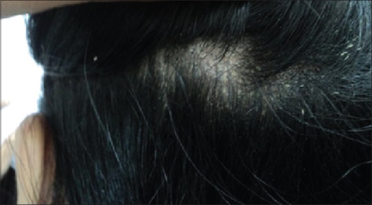
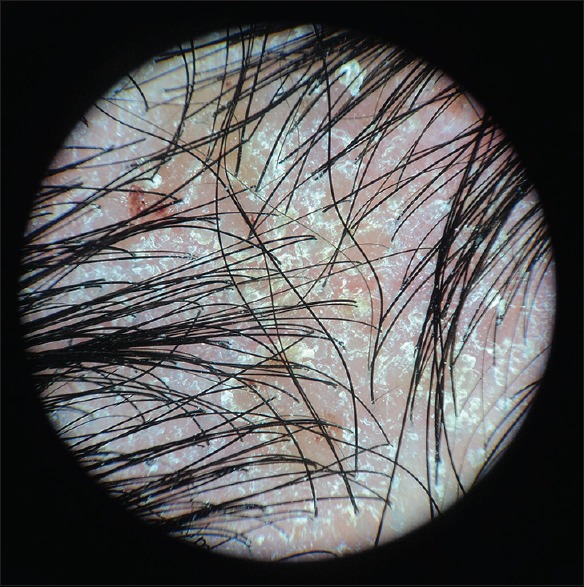
Introduction: Seborrheic dermatitis (SD) is a chronic-recidive inflammatory skin disorder with predilection in areas rich of sebaceous gland. The most common clinical manifestations are pruritus and scales. Although SD can be diagnosed without special tools, other examinations may be needed to determine additional specific therapy. Trichoscopy is one of the noninvasive tools which can help to diagnose SD as it can provide the microstructure view of the scalp.
Materials and methods: This descriptive study was conducted to explore the trichoscopic features of SD and its characteristics. There were 96 SD patients enrolled in this study. The scalp was divided into four areas, and each area was scored based on Seborrheic Area Severity Index, comprising erythema, desquamation, number of papules, and percentage of lesion area. The most severe area was examined with a trichoscopy to observe the characteristics of hair and scalp. The association between trichoscopic findings and SD severity was analyzed with Fisher's exact test.
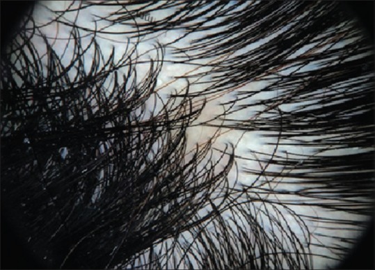
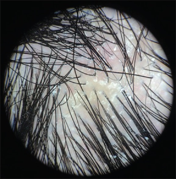
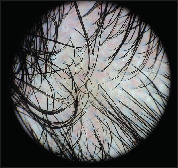
Results: Overall, the participants were 36% males and 64% females with the mean age of 30 (13-70) years old. Based on the trichoscopic examination, the most common findings were thick hair shafts (72%), white scales (69%), arborizing thin vessels (38%), yellowish area (36%), and structureless red area (19%). These findings were not significantly different between mild and moderate SD (P > 0.05).
Conclusion: Considering the merits and demerits of trichoscopic examination, it can be helpful to aid the diagnosis of SD. Further studies in Asian population with greater sample size are needed to demonstrate more significant result.
Keywords: Diagnostic tools; scalp; seborrheic dermatitis; seborrheic dermatitis area severity index; trichoscopy.
Conflict of interest statement
There are no conflicts of interest.
Figures
References
LinkOut - more resources
Full Text Sources
Other Literature Sources