Comparison of Vacuum-Assisted Closure Therapy and Conventional Dressing on Wound Healing in Patients with Diabetic Foot Ulcer: A Randomized Controlled Trial
- PMID: 31007506
- PMCID: PMC6452767
- DOI: 10.4103/njs.NJS_14_18
Comparison of Vacuum-Assisted Closure Therapy and Conventional Dressing on Wound Healing in Patients with Diabetic Foot Ulcer: A Randomized Controlled Trial
Abstract
Background: Vacuum-assisted closure (VAC) therapy has been shown to be beneficial in a variety of wounds. However, evidence of its benefit in diabetic foot ulcers (DFUs), especially with respect to Indian population, is sparse.
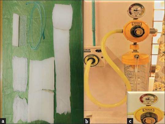
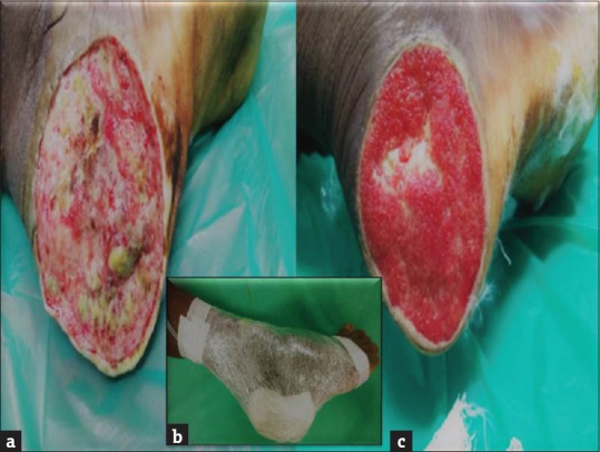
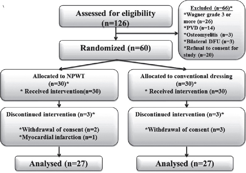
Methodology: This randomized controlled trial included DFUs of Wagner's Grades 1 and 2. Patients were further stratified with respect to DFU size <10 cm and ≥10 cm. Patients with vascular disease, osteomyelitis, and bilateral DFUs were excluded from the study. The enrolled patients were randomized to receive VAC therapy or conventional dressing. The time to wound healing, granulation tissue formation, and complications such as pain, infection, and bleeding were compared between the two groups.
Results: A total of sixty patients were randomized, of which 27 in each group were analyzed. The mean time to healing in days was significantly less in VAC group (22.52 vs. 3.85; P < 0.0001). Mean time to achieve 75%-100% granulation tissue cover was significantly less in VAC group (23.33 vs. 32.15; P < 0.0001). Rate of granulation tissue formation was also found to be significantly better in VAC group (2.91 cm2/day vs. 2.16 cm2/day; P = 0.0306). There was no difference between the two groups with respect to wound infection and bleeding which are commonly attributed to VAC therapy. VAC therapy group had significantly lesser pain at week 3 (Visual Analog Scale score 3 vs. 4; P = 0.004).
Conclusion: VAC therapy significantly decreases the time to complete wound healing, hastens granulation tissue formation, and reduces the ulcer area compared to conventional dressing. The study did not find any significant increase in the bleeding and infection in the VAC therapy group.
Keywords: Diabetic foot ulcer; granulation; negative pressure wound therapy; vacuum-assisted closure therapy; wound healing.
Conflict of interest statement
There are no conflicts of interest.
Figures
Similar articles
-
Efficacy of negative pressure wound therapy using vacuum-assisted closure combined with photon therapy for management of diabetic foot ulcers.Ther Clin Risk Manag. 2018 Oct 25;14:2113-2118. doi: 10.2147/TCRM.S164161. eCollection 2018. Ther Clin Risk Manag. 2018. PMID: 30498354 Free PMC article.
-
Evaluation of low-cost custom made VAC therapy compared with conventional wound dressings in the treatment of non-healing lower limb ulcers in lower socio-economic group patients of Kashmir valley.J Orthop Surg Res. 2015 Dec 10;10:183. doi: 10.1186/s13018-015-0314-5. J Orthop Surg Res. 2015. Retraction in: J Orthop Surg Res. 2017 Apr 20;12(1):62. doi: 10.1186/s13018-017-0558-3. PMID: 26654318 Free PMC article. Retracted.
-
Effectiveness of vacuum-assisted closure (VAC) therapy in the healing of chronic diabetic foot ulcers.Ann Acad Med Singap. 2010 May;39(5):353-8. Ann Acad Med Singap. 2010. PMID: 20535423
-
Vacuum assisted closure (VAC)/negative pressure wound therapy (NPWT) for difficult wounds: A review.J Clin Orthop Trauma. 2019 Sep-Oct;10(5):845-848. doi: 10.1016/j.jcot.2019.06.015. Epub 2019 Jun 20. J Clin Orthop Trauma. 2019. PMID: 31528055 Free PMC article. Review.
-
[Vacuum assisted wound closure --overview of lesson and applications].Cas Lek Cesk. 2006;145(9):702-7; discussion 707. Cas Lek Cesk. 2006. PMID: 17091725 Review. Slovak.
Cited by
-
Postoperative negative pressure wound therapy is associated with decreased surgical site infections in all lower extremity amputations.J Orthop. 2020 Sep 8;21:507-511. doi: 10.1016/j.jor.2020.09.005. eCollection 2020 Sep-Oct. J Orthop. 2020. PMID: 32999539 Free PMC article.
-
Effect of negative pressure wound therapy on wound thermometry in diabetic foot ulcers.J Family Med Prim Care. 2022 Nov;11(11):7001-7007. doi: 10.4103/jfmpc.jfmpc_72_22. Epub 2022 Dec 16. J Family Med Prim Care. 2022. PMID: 36993063 Free PMC article.
-
Role of nuclear factor erythroid 2-related factor 2 in negative pressure wound therapy for diabetic foot ulcers.World J Diabetes. 2025 May 15;16(5):104350. doi: 10.4239/wjd.v16.i5.104350. World J Diabetes. 2025. PMID: 40487630 Free PMC article.
-
The effect of negative pressure wound therapy on the outcome of diabetic foot ulcers: A meta-analysis.Int Wound J. 2024 Apr;21(4):e14886. doi: 10.1111/iwj.14886. Int Wound J. 2024. Retraction in: Int Wound J. 2024 Nov;21(11):e70133. doi: 10.1111/iwj.70133. PMID: 38651532 Free PMC article. Retracted.
-
Comparative study of various dressing techniques in diabetic foot ulcers in the Indian population: a single-center experience.Int J Diabetes Dev Ctries. 2023 Jan 4:1-7. doi: 10.1007/s13410-022-01163-3. Online ahead of print. Int J Diabetes Dev Ctries. 2023. PMID: 36619905 Free PMC article.
References
-
- Viswanathan V. The diabetic foot: Perspectives from Chennai, South India. Int J Low Extrem Wounds. 2007;6:34–6. - PubMed
-
- Vijay V, Narasimham DV, Seena R, Snehalatha C, Ramachandran A. Clinical profile of diabetic foot infections in South India – A retrospective study. Diabet Med. 2000;17:215–8. - PubMed
-
- Ibrahim A. IDF clinical practice recommendation on the diabetic foot: A guide for healthcare professionals. Diabetes Res Clin Pract. 2017;127:285–7. - PubMed
LinkOut - more resources
Full Text Sources
Medical