. 2019 Feb;12(1):69-84.
doi: 10.1007/s12195-018-0547-6. Epub 2018 Aug 16.

Degradation and Remodeling of Epitaxially Grown Collagen Fibrils

Affiliations

Degradation and Remodeling of Epitaxially Grown Collagen Fibrils

Juan Wang et al. Cell Mol Bioeng. 2019 Feb.

Abstract

Introduction—: The extracellular matrix (ECM) in the tumor microenvironment contains high densities of collagen that are highly aligned, resulting in directional migration called contact guidance that facilitates efficient migration out of the tumor. Cancer cells can remodel the ECM through traction force controlled by myosin contractility or proteolytic activity controlled by matrix metalloproteinase (MMP) activity, leading to either enhanced or diminished contact guidance.

Methods—: Recently, we have leveraged the ability of mica to epitaxially grow aligned collagen fibrils in order to assess contact guidance. In this article, we probe the mechanisms of remodeling of aligned collagen fibrils on mica by breast cancer cells.

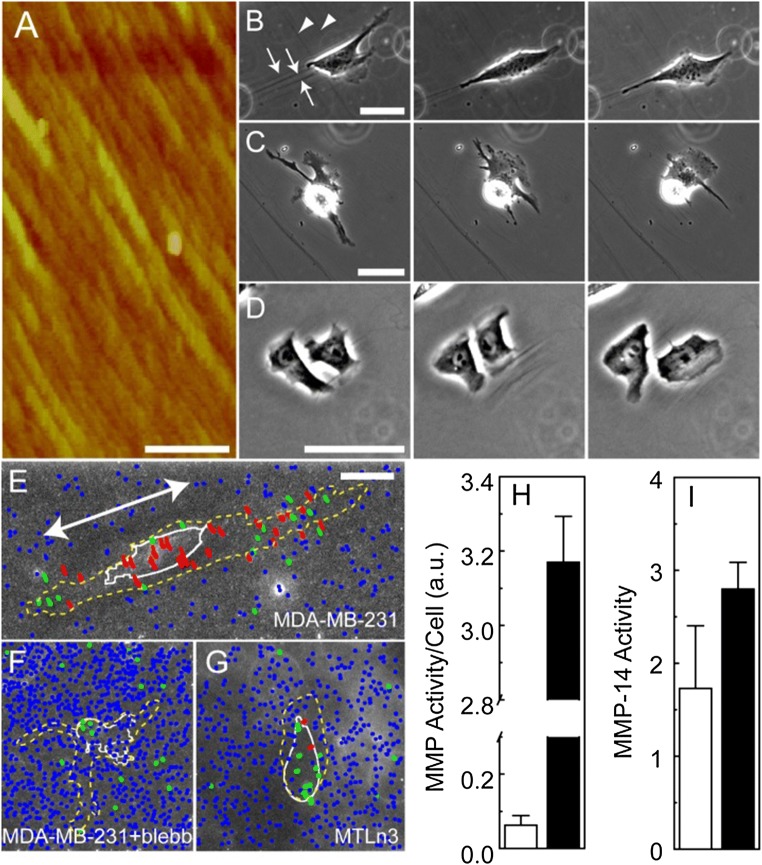
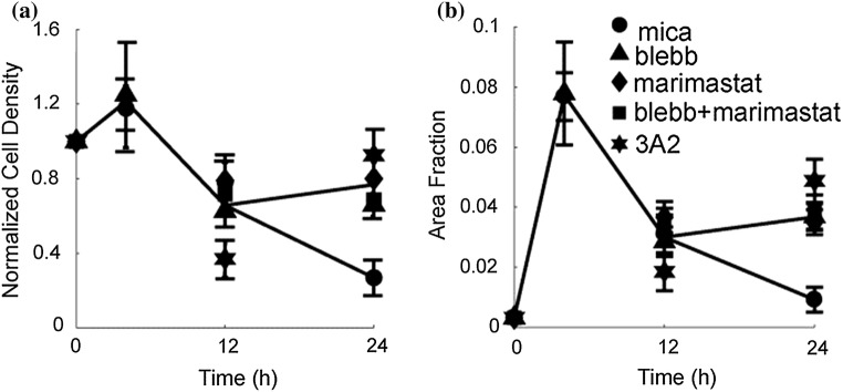
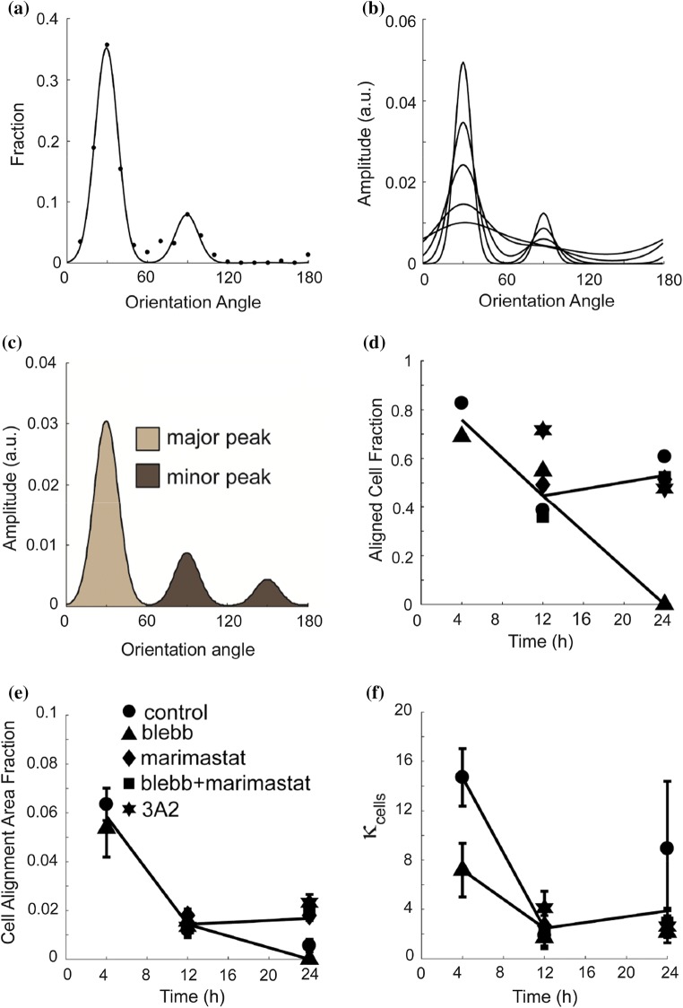
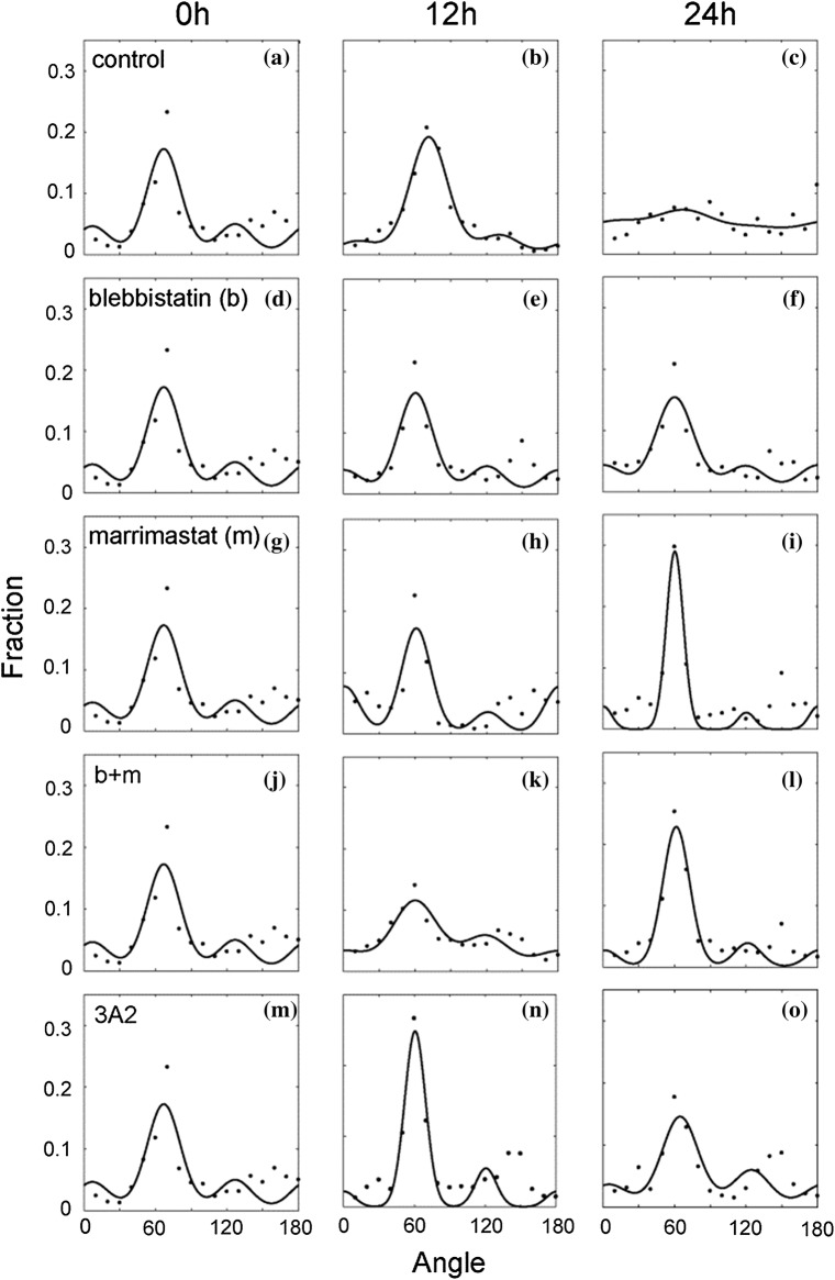
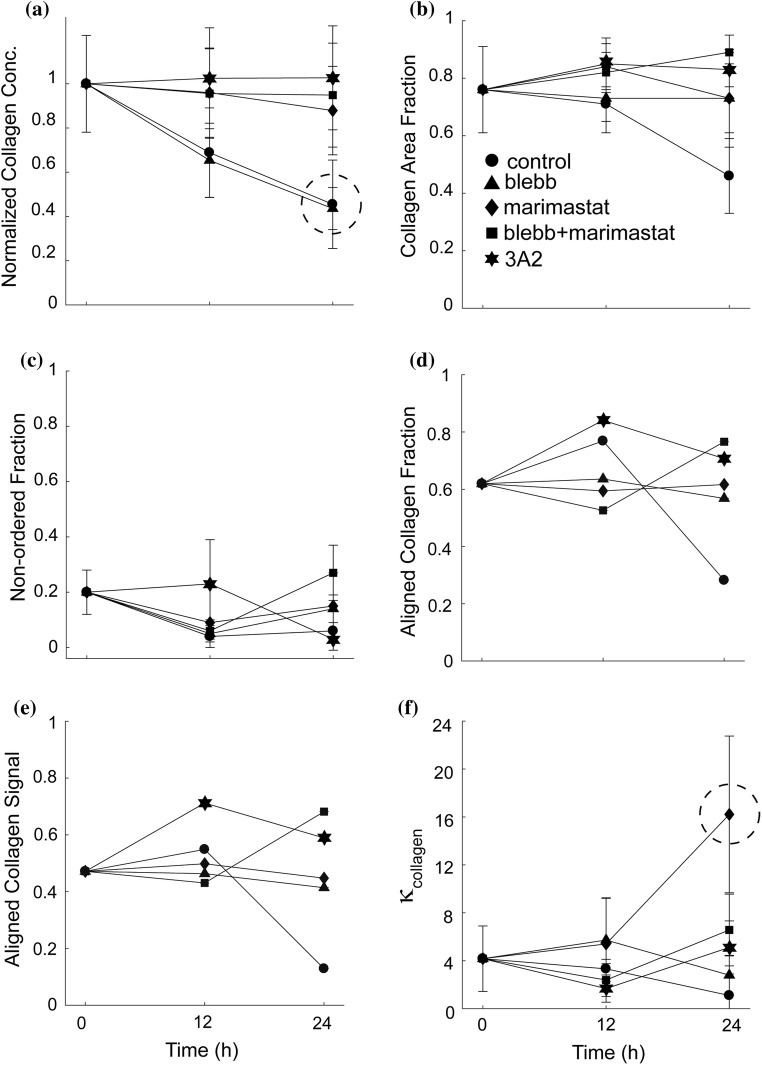
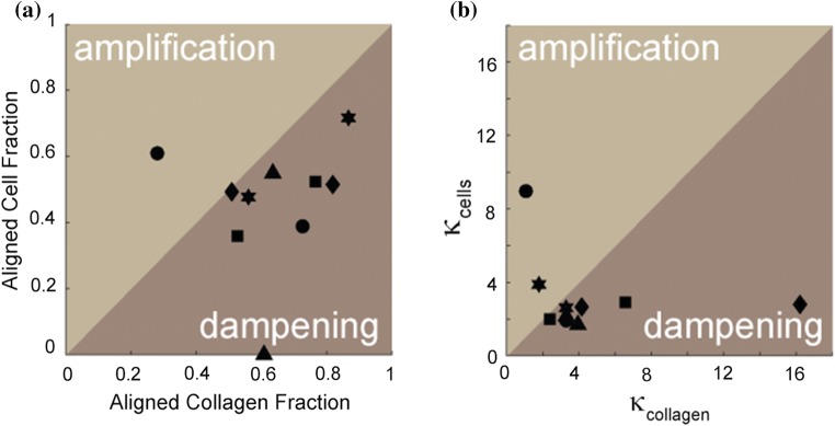
Results—: We show that cells that contact guide with high fidelity (MDA-MB-231 cells) exert more force on the underlying collagen fibrils than do cells that contact guide with low fidelity (MTLn3 cells). These high traction cells (MDA-MB-231 cells) remodel collagen fibrils over hours, pulling so hard that the collagen fibrils detach from the surface, effectively delaminating the entire contact guidance cue. Myosin or MMP inhibition decreases this effect. Interestingly, blocking MMP appears to increase the alignment of cells on these substrates, potentially allowing the alignment through myosin contractility to be uninhibited. Finally, amplification or dampening of contact guidance with respect to a particular collagen fibril organization is seen under different conditions.

Conclusions—: Both myosin II contractility and MMP activity allow MDA-MB-231 cells to remodel and eventually destroy epitaxially grown aligned collagen fibrils.

Keywords: Directed migration; Function blocking antibody; MMP-14; MT1-MMP; Second harmonic generation.


Conflict of interest statement

CONFLICT OF INTEREST Juan Wang, Anuraag Boddupalli, Joseph Koelbl, Dong Hyun Nam, Xin Ge, Kaitlin M. Bratlie and Ian C. Schneider state they have no conflicts of interest.

Figures

Figure 1
Figure 1
Cell alignment and traction on epitaxially grown aligned collagen fibrils. (a) Atomic force microscopy image of epitaxially grown, aligned collagen fibrils. The scale bar is 1 μm. (bd) Timelapse phase contrast images of an (a) MDA-MB-231 cell, (b) MDA-MB-231 cell treated with blebbistatin and (c) MTLn3 cell on aligned collagen fibrils. (a) Collagen fibrils are marked with arrows and move between 0 min (left), 200 min (center) and 400 min (right). Arrow head marks scratches in the mica that do not deform over time. The scale bar length is 30 μm. Trajectories of fluorescent nanospheres adsorbed on aligned collagen fibrils assembled on mica for (e) MDA-MB-231 cells, (f) MDA-MB-231 cells treated with blebbistatin (30 μM) and (g) MTLn3 cells. Beads are colored differently depending on their mean displacement. Red indicates mean displacements larger than 600 nm, green indicates mean displacements between 300 and 600 nm and blue represents mean displacement smaller than 300 nm. The cell outline from one time point is shown in white and the region over which the cell migrates over the entire time lapse is shown as the yellow dashed line. The primary migration direction for (e) is shown as the arrow. (h) MMP activity in MDA-MB-231 (white) and MTLn3 (black) cells MMP activity on a per cell basis. Note the break in the axis. (i) Activity of the head domain of MMP-14 in the media used for MDA-MB-231 (white) and MTLn3 (black) cell migration experiments. Error bars are 95% confidence intervals.
Figure 2
Figure 2
(a) Normalized cell density and (b) area fraction occupied by MDA-MB-231 cells change with time on aligned collagen fibrils assembled on mica (circles). The decrease seen at 24 h is blocked when treated with a myosin II-mediated contractility inhibitor (blebbistatin), a pan-MMP inhibitor (marimastat) or an MMP-14 function blocking antibody (3A2). Blebbistatin (30 μM, triangle), marimastat (10 μM, diamond), marimastat and blebbistatin (10 and 30 μM, squares) or 3A2 antibody (500 μM, star). Error bars represent 95% confidence intervals. Nexperiments > 3 and Ncell > 160.
Figure 3
Figure 3
Cell alignment of MDA-MB-231 cells treated with a myosin II-mediated contractility inhibitor (blebbistatin), a pan-MMP inhibitor (marimastat) or an MMP-14 function blocking antibody (3A2). (a) The angle distribution of cells on aligned collagen fibrils assembled on mica at 4 h (circles). The sum of three von Mises distributions separated by 60 degrees was used to fit the data. (b) The sum of three von Mises distributions with different values for the distribution spread parameter (κcells=1, 2, 5, 10 and 20). As κcells increases, the spread of the distribution descreases. κcells is inversely proportional to the full width half max. (c) Schematic of cell angle distribution for the one major and two minor peaks. The cell alignment fraction is calculated as fraction of the total area that is under the major peak. (d) Cell alignment fraction, (e) cell alignment area fraction and (f) distribution spread parameter, κcells as a function of time and exposed to different inhibitors. Cells plated on aligned collagen fibrils assembled on mica (control, circle) treated with blebbistatin (30 μM, triangle), marimastat (10 μM, diamond), marimastat and blebbistatin (30 and 10 μM, squares) or 3A2 antibody (500 nM, star). Error bars represent 95% confidence intervals. Nexperiments > 3 and Ncell > 160.
Figure 4
Figure 4
Collagen alignment on mica substrates seeded with MDA-MB-231 cells treated with a myosin II-mediated contractility inhibitor (blebbistatin), a pan-MMP inhibitor (marimastat) or an MMP-14 function blocking antibody (3A2). The angular distribution of collagen of MDA-MB-231 cells at 0, 12, and 24 h treated with (ac) no inhibitor (df) blebbistatin (gi) marimastat (jl) both marimastat and blebbistatin, and (mo) 3A2. Circles are data points and the lines are the von Mises fits to the data. Nexperiments > 3 and Nimages > 9.
Figure 5
Figure 5
Collagen fiber alignment on mica substrates seeded with MDA-MB-231 cells treated with a myosin II-mediated contractility inhibitor (blebbistatin), a pan-MMP inhibitor (marimastat) or an MMP-14 function blocking antibody (3A2). (a) The relative intensity of the collagen fibers on the mica-based substrates over 24 h, normalized to the initial intensity of the control at 0 h. The mica and blebbistatin substrates are circled. (b) The collagen area fraction of the sum of both ordered and non-ordered collagen fractions. (c) The non-ordered collagen fractions of the collagen signal. (d) Collagen alignment fraction, (e) Aligned collagen signal fraction, as a product of the aligned collagen fraction and the normalized collagen intensity and (f) distribution spread parameter, κcollagen. Cells plated on aligned collagen fibrils assembled on mica (control, circle) treated with blebbistatin (30 μM, triangle), marimastat (10 μM, diamond), marimastat and blebbistatin (10 and 30 μM, squares) or 3A2 antibody (500 nM, star). Error bars represent 95% confidence intervals. Nexperiments > 3 and Nimages > 9.
Figure 6
Figure 6
(a) Aligned cell and collagen fraction and (b) distribution spread parameter (κcollagen) for both cells and collagen under control and inhibitor conditions. Cells plated on aligned collagen fibrils assembled on mica (circle) treated with blebbistatin (30 μM, triangle), marimastat (10 μM, diamond), marimastat and blebbistatin (10 and 30 μM, squares) or 3A2 antibody (500 nM, star). Cell amplification of the directional cue occurs when either the alignment fraction or the κcollagen is larger for cells than for collagen. Cell dampening of the directional cue occurs when either the alignment fraction or the κcollagen is smaller for cells than for collagen.

Similar articles

Cited by

References

    1. Alexander NR, Branch KM, Parekh A, Clark ES, Lwueke LC, Guelcher SA, Weaver AM. Extracellular matrix rigidity promotes invadopodia activity. Curr. Biol. 2008;18:1295–1299. doi: 10.1016/j.cub.2008.07.090. - DOI - PMC - PubMed
    1. Barocas VH, Tranquillo RT. An anisotropic biphasic theory of tissue-equivalent mechanics: the interplay among cell traction, fibrillar network deformation, fibril alignment, and cell contact guidance. J. Biomech. Eng. Trans. ASME. 1997;119:137–145. doi: 10.1115/1.2796072. - DOI - PubMed
    1. Biela SA, Su Y, Spatz JP, Kemkemer R. Different sensitivity of human endothelial cells, smooth muscle cells and fibroblasts to topography in the nano-micro range. Acta Biomater. 2009;5:2460–2466. doi: 10.1016/j.actbio.2009.04.003. - DOI - PubMed
    1. Chiu C-L, Digman MA, Gratton E. Cell matrix remodeling ability shown by image spatial correlation. J. Biophys. 2013;2013:8. doi: 10.1155/2013/532030. - DOI - PMC - PubMed
    1. Clark P, Connolly P, Curtis ASG, Dow JAT, Wilkinson CDW. Topographical control of cell behavior. 2. Multiple grooved substrata. Development. 1990;108:635–644. - PubMed

LinkOut - more resources