

Amyloid Peptide β 1-42 Induces Integrin α IIb β 3 Activation, Platelet Adhesion, and Thrombus Formation in a NADPH Oxidase-Dependent Manner
- PMID: 31007831
- PMCID: PMC6441506
- DOI: 10.1155/2019/1050476
Amyloid Peptide β 1-42 Induces Integrin α IIb β 3 Activation, Platelet Adhesion, and Thrombus Formation in a NADPH Oxidase-Dependent Manner
Abstract
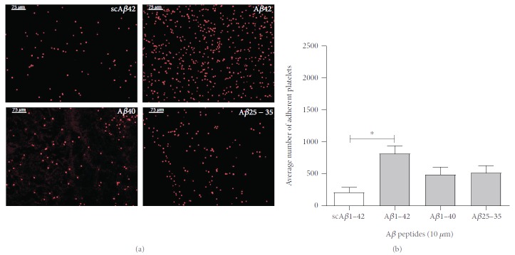
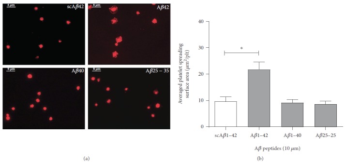
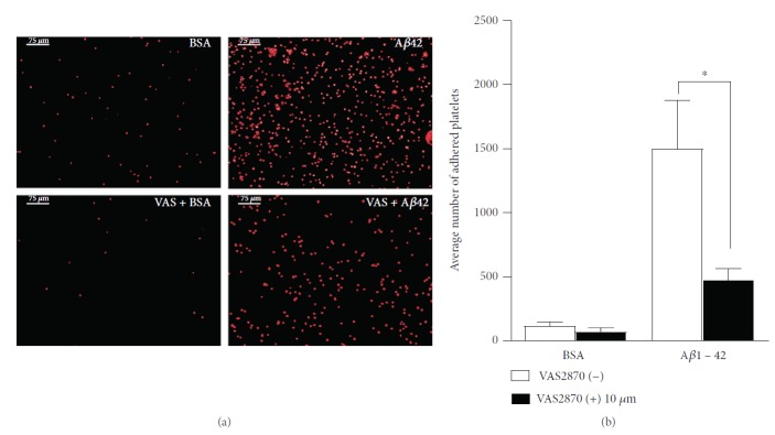
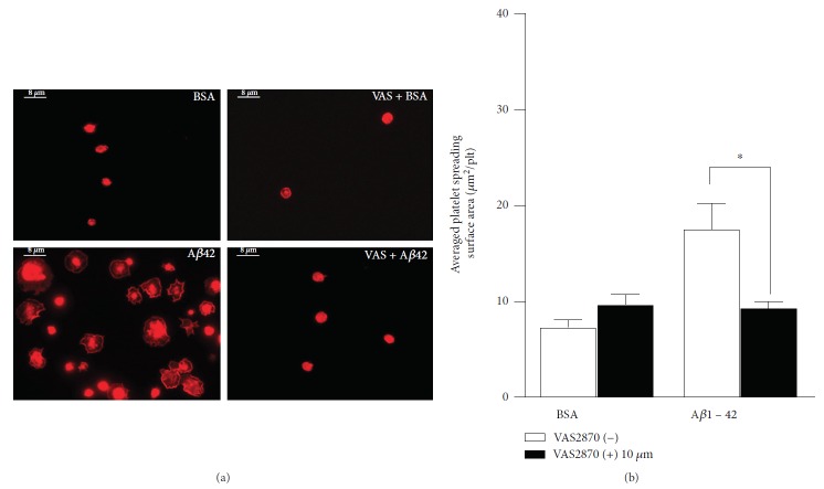
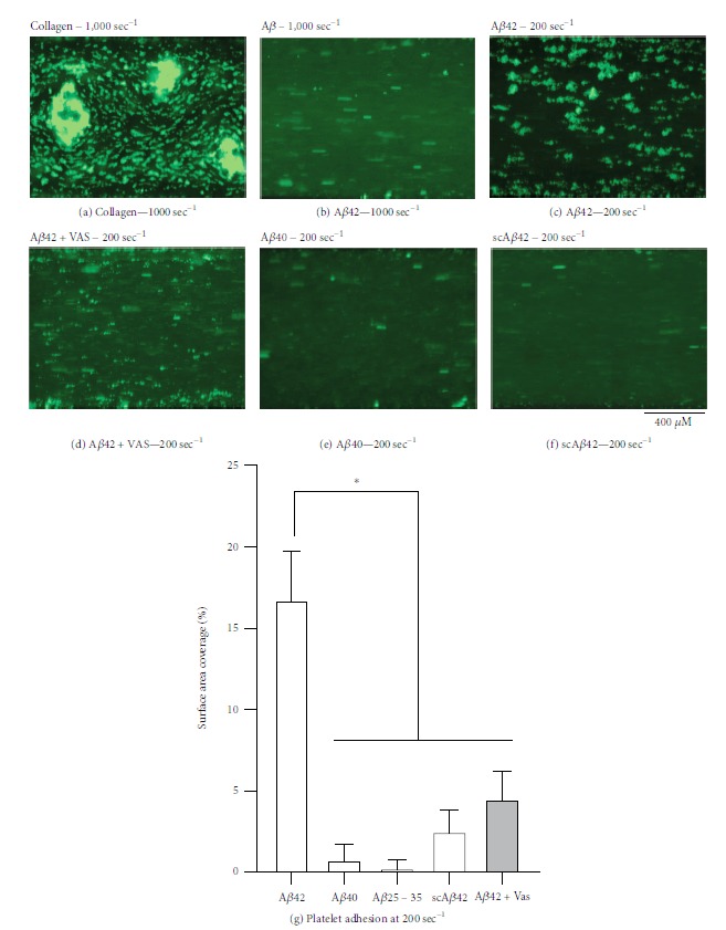
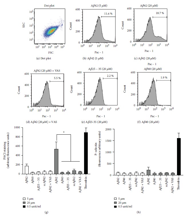
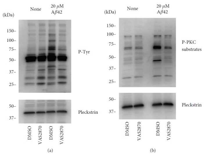
The progression of Alzheimer's dementia is associated with neurovasculature impairment, which includes inflammation, microthromboses, and reduced cerebral blood flow. Here, we investigate the effects of β amyloid peptides on the function of platelets, the cells driving haemostasis. Amyloid peptide β1-42 (Aβ1-42), Aβ1-40, and Aβ25-35 were tested in static adhesion experiments, and it was found that platelets preferentially adhere to Aβ1-42 compared to other Aβ peptides. In addition, significant platelet spreading was observed over Aβ1-42, while Aβ1-40, Aβ25-35, and the scAβ1-42 control did not seem to induce any platelet spreading, which suggested that only Aβ1-42 activates platelet signalling in our experimental conditions. Aβ1-42 also induced significant platelet adhesion and thrombus formation in whole blood under venous flow condition, while other Aβ peptides did not. The molecular mechanism of Aβ1-42 was investigated by flow cytometry, which revealed that this peptide induces a significant activation of integrin αIIbβ3, but does not induce platelet degranulation (as measured by P-selectin membrane translocation). Finally, Aβ1-42 treatment of human platelets led to detectable levels of protein kinase C (PKC) activation and tyrosine phosphorylation, which are hallmarks of platelet signalling. Interestingly, the NADPH oxidase (NOX) inhibitor VAS2870 completely abolished Aβ1-42-dependent platelet adhesion in static conditions, thrombus formation in physiological flow conditions, integrin αIIbβ3 activation, and tyrosine- and PKC-dependent platelet signalling. In summary, this study highlights the importance of NOXs in the activation of platelets in response to amyloid peptide β1-42. The molecular mechanisms described in this manuscript may play an important role in the neurovascular impairment observed in Alzheimer's patients.
Figures
Comment in
-
Oxidative Stress and Mitochondrial Damage in Neurodegenerative Diseases: From Molecular Mechanisms to Targeted Therapies.Oxid Med Cell Longev. 2020 May 4;2020:1270256. doi: 10.1155/2020/1270256. eCollection 2020. Oxid Med Cell Longev. 2020. PMID: 32454930 Free PMC article. No abstract available.
Similar articles
-
The novel NOX inhibitor 2-acetylphenothiazine impairs collagen-dependent thrombus formation in a GPVI-dependent manner.Br J Pharmacol. 2013 Jan;168(1):212-24. doi: 10.1111/j.1476-5381.2012.02130.x. Br J Pharmacol. 2013. PMID: 22881838 Free PMC article.
-
Amyloid precursor protein is required for in vitro platelet adhesion to amyloid peptides and potentiation of thrombus formation.Cell Signal. 2018 Dec;52:95-102. doi: 10.1016/j.cellsig.2018.08.017. Epub 2018 Aug 30. Cell Signal. 2018. PMID: 30172024
-
A novel flow cytometry assay using dihydroethidium as redox-sensitive probe reveals NADPH oxidase-dependent generation of superoxide anion in human platelets exposed to amyloid peptide β.Platelets. 2019;30(2):181-189. doi: 10.1080/09537104.2017.1392497. Epub 2017 Dec 5. Platelets. 2019. PMID: 29206074
-
Relevance of N-terminal residues for amyloid-β binding to platelet integrin αIIbβ3, integrin outside-in signaling and amyloid-β fibril formation.Cell Signal. 2018 Oct;50:121-130. doi: 10.1016/j.cellsig.2018.06.015. Epub 2018 Jun 30. Cell Signal. 2018. PMID: 29964150
-
Platelet Signaling in Primary Haemostasis and Arterial Thrombus Formation: Part 2.Hamostaseologie. 2018 Nov;38(4):211-222. doi: 10.1055/s-0038-1675149. Epub 2018 Nov 30. Hamostaseologie. 2018. PMID: 30500969 Review.
Cited by
-
NADPH Oxidases Are Required for Full Platelet Activation In Vitro and Thrombosis In Vivo but Dispensable for Plasma Coagulation and Hemostasis.Arterioscler Thromb Vasc Biol. 2021 Feb;41(2):683-697. doi: 10.1161/ATVBAHA.120.315565. Epub 2020 Dec 3. Arterioscler Thromb Vasc Biol. 2021. PMID: 33267663 Free PMC article.
-
Role of Thrombosis in Neurodegenerative Diseases: An Intricate Mechanism of Neurovascular Complications.Mol Neurobiol. 2025 Apr;62(4):4802-4836. doi: 10.1007/s12035-024-04589-4. Epub 2024 Nov 1. Mol Neurobiol. 2025. PMID: 39482419 Review.
-
Bleeding is increased in amyloid precursor protein knockout mouse.Res Pract Thromb Haemost. 2020 Jun 14;4(5):823-828. doi: 10.1002/rth2.12375. eCollection 2020 Jul. Res Pract Thromb Haemost. 2020. PMID: 32685890 Free PMC article.
-
Alzheimer's Disease-Thrombosis Comorbidity: A Growing Body of Evidence from Patients and Animal Models.Cells. 2025 Jul 12;14(14):1069. doi: 10.3390/cells14141069. Cells. 2025. PMID: 40710322 Free PMC article. Review.
-
Reversal of β-Amyloid-Induced Microglial Toxicity In Vitro by Activation of Fpr2/3.Oxid Med Cell Longev. 2020 Jun 13;2020:2139192. doi: 10.1155/2020/2139192. eCollection 2020. Oxid Med Cell Longev. 2020. PMID: 32617132 Free PMC article.
References
MeSH terms
Substances
Grants and funding
LinkOut - more resources
Full Text Sources
Medical
