

Blood Flow Assessment of Arteriovenous Malformations Using Intraoperative Indocyanine Green Videoangiography
- PMID: 31007890
- PMCID: PMC6441520
- DOI: 10.1155/2019/7292304
Blood Flow Assessment of Arteriovenous Malformations Using Intraoperative Indocyanine Green Videoangiography
Abstract
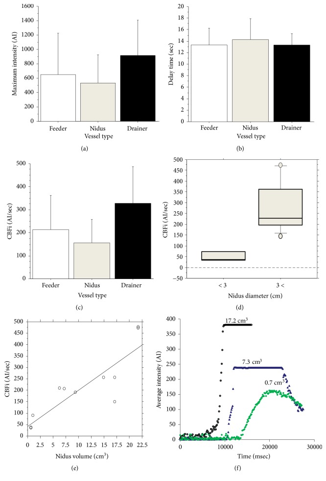
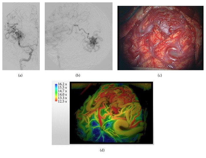
Intraoperative indocyanine green (ICG) videoangiography is widely used in patients undergoing neurosurgery. FLOW800 is a recently developed analytical tool for ICG videoangiography to assess semi-quantitative flow dynamics; however, its efficacy is unknown. In this study, we evaluated its functionality in the assessment of flow dynamics of arteriovenous malformation (AVM) through ICG videoangiography under clinical settings. ICG videoangiography was performed in the exposed AVM in eight patients undergoing surgery. FLOW800 analysis was applied directly, and gray-scale and color-coded maps of the surgical field were obtained. After surgery, a region of interest was placed on the individual vessels to obtain time-intensity curves. Parameters of flow dynamics, including the maximum intensity, transit time, and cerebral blood flow index, were calculated using the curves. The color-coded maps provided high-resolution images; however, reconstruction of colored images was restricted by the depth, approach angle, and brain swelling. Semi-quantitative parameters were similar among the feeders, niduses, and drainers. However, a higher cerebral blood flow index was observed in the feeders of large AVM (>3 cm) than in those of small AVM (P < 0.05). Similarly, the cerebral blood flow index values were positively correlated with the nidus volume (P < 0.01). FLOW800 enabled visualization of the AVM structure and safer resection, except in case of deep-seated AVM. Moreover, semi-quantitative values in the individual vessels through using ICG intensity diagram showed different patterns according to size of the AVM. ICG videoangiography showed good performance in evaluating flow dynamics of the AVM in patients undergoing surgery.
Figures


Similar articles
-
Uses and limitations of indocyanine green videoangiography for flow analysis in arteriovenous malformation surgery.J Clin Neurosci. 2013 Feb;20(2):224-32. doi: 10.1016/j.jocn.2011.12.038. Epub 2012 Dec 29. J Clin Neurosci. 2013. PMID: 23277126
-
Intraoperative Anatomical and Hemodynamic Analysis of Intracerebral Arteriovenous Malformations by Semi-quantitative Color-coded Indocyanine Green Videoangiography.Asian J Neurosurg. 2017 Oct-Dec;12(4):638-643. doi: 10.4103/ajns.AJNS_62_14. Asian J Neurosurg. 2017. PMID: 29114275 Free PMC article.
-
Efficacy of intraarterial superselective indocyanine green videoangiography in cerebral arteriovenous malformation surgery in a hybrid operating room.J Neurosurg. 2020 May 22;134(5):1544-1552. doi: 10.3171/2020.3.JNS20319. Print 2021 May 1. J Neurosurg. 2020. PMID: 32442970
-
Usefulness of tumor blood flow imaging by intraoperative indocyanine green videoangiography in hemangioblastoma surgery.World Neurosurg. 2014 Sep-Oct;82(3-4):e495-501. doi: 10.1016/j.wneu.2013.02.009. Epub 2013 Feb 8. World Neurosurg. 2014. PMID: 23396070 Review.
-
Role of intraoperative indocyanine green video-angiography to identify small, posterior fossa arteriovenous malformations mimicking cavernous angiomas. Technical report and review of the literature on common features of these cerebral vascular malformations.Clin Neurol Neurosurg. 2015 Nov;138:45-51. doi: 10.1016/j.clineuro.2015.07.016. Epub 2015 Jul 26. Clin Neurol Neurosurg. 2015. PMID: 26276727 Review.
Cited by
-
Non-Angry Superficial Draining Veins: A New Technique in Identifying the Extent of Nidus Excision during Cerebral Arteriovenous Malformation Surgery.Brain Sci. 2023 Feb 20;13(2):366. doi: 10.3390/brainsci13020366. Brain Sci. 2023. PMID: 36831909 Free PMC article.
-
Pediatric Low-Grade Glioma Surgery with Sodium Fluorescein: Efficient Localization for Removal and Association with Intraoperative Pathological Sampling.Diagnostics (Basel). 2022 Nov 23;12(12):2927. doi: 10.3390/diagnostics12122927. Diagnostics (Basel). 2022. PMID: 36552934 Free PMC article.
-
Application of Indocyanine Green During Arteriovenous Malformation Surgery: Evidence, Techniques, and Practical Pearls.Front Surg. 2019 Dec 11;6:70. doi: 10.3389/fsurg.2019.00070. eCollection 2019. Front Surg. 2019. PMID: 31921884 Free PMC article. Review.
-
Microvascular Cortical Dynamics in Minimal Invasive Deep-Seated Brain Tumour Surgery.Cancers (Basel). 2025 Apr 22;17(9):1392. doi: 10.3390/cancers17091392. Cancers (Basel). 2025. PMID: 40361321 Free PMC article.
-
Continuous blood flow visualization with laser speckle contrast imaging during neurovascular surgery.Neurophotonics. 2022 Apr;9(2):021908. doi: 10.1117/1.NPh.9.2.021908. Epub 2022 Mar 7. Neurophotonics. 2022. PMID: 35265733 Free PMC article.
References
-
- Bekelis K., Missios S., Desai A., Eskey C., Erkmen K. Magnetic resonance imaging/magnetic resonance angiography fusion technique for intraoperative navigation during microsurgical resection of cerebral arteriovenous malformations. Neurosurgical Focus. 2012;32(5, article E7) - PubMed
-
- Killory B. D., Nakaji P., Maughan P. H., Wait S. D., Spetzler R. F. Evaluation of angiographically occult spinal dural arteriovenous fistulae with surgical microscope-integrated intraoperative near-infrared indocyanine green angiography: Report of 3 cases. Neurosurgery. 2011;68(3):781–787. doi: 10.1227/NEU.0b013e318207ac3b. - DOI - PubMed
LinkOut - more resources
Full Text Sources
