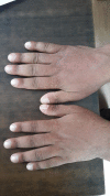
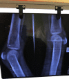
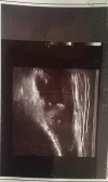
Inflammatory variant of pachydermoperiostosis responding to methotrexate: a report of two cases
- PMID: 31007935
- PMCID: PMC6467096
- DOI: 10.1093/omcr/omy128
Inflammatory variant of pachydermoperiostosis responding to methotrexate: a report of two cases
Abstract
Pachydermoperiostosis is a rare genetic disorder characterized by skin thickening, digital clubbing and periostitis. The pathogenesis is incompletely understood and there are no proven treatments for its manifestations. Although arthritis has been reported in 20-40% cases, most are non-inflammatory in nature and usually treated symptomatically with steroids or NSAIDs. We report two cases of pachydermoperiostosis with inflammatory variant of arthritis and raised inflammatory markers who were treated with tapering dose of prednisolone for 6 weeks and maintained on long-term low dose methotrexate like rheumatoid arthritis and followed for 2 years. In both cases, methotrexate was well tolerated and helped in maintaining symptomatic improvement and slowed the disease progression with significant steroid and NSAID sparing effect. We concluded that there exists an inflammatory subtype of disease where methotrexate can be beneficial.
Figures


Similar articles
-
[Primary hypertrophic osteoarthropathy (pachydermoperiostosis). Report of two familial cases and literature review].Reumatol Clin. 2009 Nov-Dec;5(6):259-63. doi: 10.1016/j.reuma.2009.01.007. Epub 2009 Sep 26. Reumatol Clin. 2009. PMID: 21794626 Spanish.
-
Pachydermoperiostosis in a patient with chronic hepatitis B virus infection referred as acromegaly: a case report.J Med Case Rep. 2018 Mar 8;12(1):59. doi: 10.1186/s13256-018-1578-2. J Med Case Rep. 2018. PMID: 29514715 Free PMC article.
-
A complicated case of pachydermoperiostosis with spondyloarthritides: a case report.J Med Case Rep. 2013 Dec 13;7:268. doi: 10.1186/1752-1947-7-268. J Med Case Rep. 2013. PMID: 24330652 Free PMC article.
-
[Methotrexate and non-steroidal anti-inflammatory agent combination in rheumatoid arthritis].Therapie. 1997 Mar-Apr;52(2):133-7. Therapie. 1997. PMID: 9231508 Review. French.
-
Leflunomide: a review of its use in active rheumatoid arthritis.Drugs. 1999 Dec;58(6):1137-64. doi: 10.2165/00003495-199958060-00010. Drugs. 1999. PMID: 10651393 Review.
Cited by
-
Comprehensive Treatment of a Rare Case of Complete Primary Pachydermoperiostosis with Large Facial Keloid Scars: A Case Report and Literature Review.Case Rep Dermatol. 2024 Mar 4;16(1):63-69. doi: 10.1159/000536550. eCollection 2024 Jan-Dec. Case Rep Dermatol. 2024. PMID: 38440721 Free PMC article.
References
-
- Carcassi U. History of hypertrophic osteoarthropathy (HOA). Clin Exp Rheumatol 1992;10:3–7. - PubMed
-
- Reginato AJ, Shipachasse V, Guerrero R. Familial idiopathic hypertrophic osteoarthropathy and cranial suture defects in children. Skeletal Radiol 1982;8:105–9. - PubMed
-
- Castori M, Sinibaldi L, Mingarelli R, Lachman RS, Rimoin DL, Dallapiccola B. Pachydermoperiostosis: an update. Clin Genet 2005;68:477–86. - PubMed
-
- Uppal S, Diggle CP, Carr IM, Fishwick CW, Ahmed M, Ibrahim GH, et al. . Mutations in 15-hydroxyprostaglandin dehydrogenase cause primary hypertrophic osteoarthropathy. Nat Genet 2008;40:789–93. - PubMed
-
- Martinez-Lavin M. Pathogenesis of hypertrophic osteoarthropathy. Clin Exp Rheumatol 1992;10:49–50. - PubMed
Publication types
LinkOut - more resources
Full Text Sources

