

Intramural duodenal hematoma: clinical course and imaging findings
- PMID: 31007947
- PMCID: PMC6456848
- DOI: 10.1177/2058460119836256
Intramural duodenal hematoma: clinical course and imaging findings
Abstract
Background: Intramural duodenal hematoma is a rare condition. Different imaging modalities are at hand for diagnosis.
Purpose: To identify patients with intramural duodenal hematoma and report imaging findings and clinical courses.
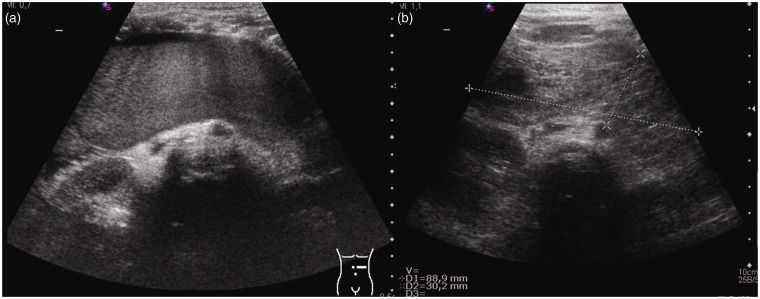
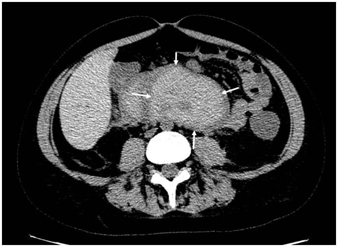
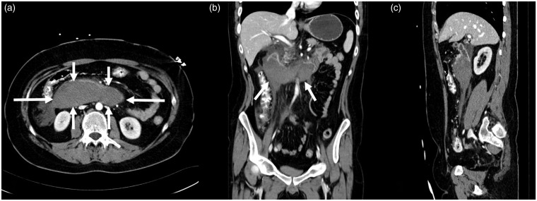
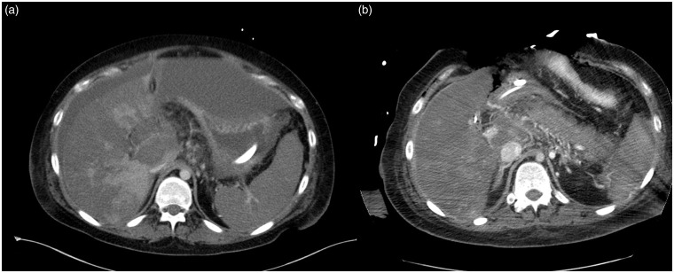
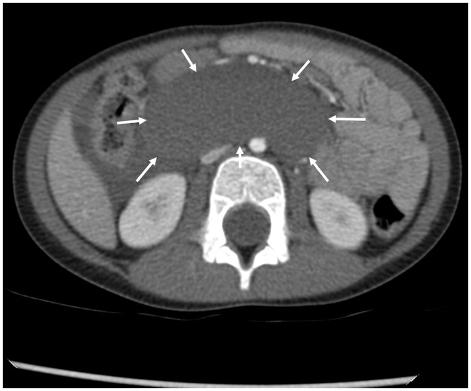
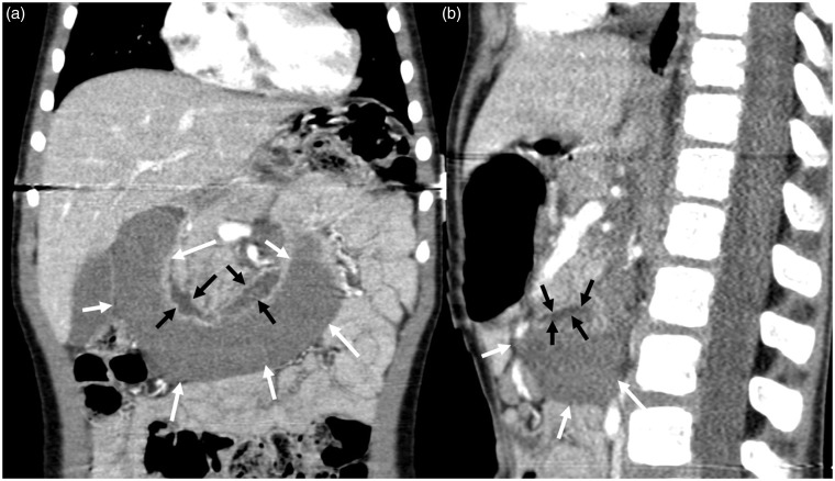
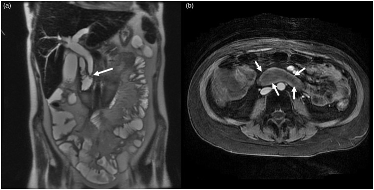
Material and methods: Typical imaging patterns using ultrasound, computed tomography, and magnetic resonance imaging were carried out on 10 patients.
Results: The mean patient age was 7.5 years. The average disease duration was 13 months. Clinical signs of improvement were observed within 16 days. Residues were still detectable at long-term follow-up.
Conclusion: For patients with intramural duodenal wall hematoma, diagnosis should be considered early. Typical imaging findings should be known to ensure optimal treatment.
Keywords: Abdomen/GI; adults and pediatrics; computed tomography; hemorrhage; small bowel; ultrasound.
Figures
References
-
- Li Y, Fan Z, Xu L, et al. Prospective ECG-gated 320-row CT angiography of the whole aorta and coronary arteries. Eur Radiol 2012; 22:2432–2440. - PubMed
-
- Ahn SS, Kim BM, Suh SH, et al. Spontaneous symptomatic intracranial vertebrobasilar dissection: initial and follow-up imaging findings. Radiology 2012; 264:196–202. - PubMed
-
- Costelloe J, Mc Cormack O, Reynolds JV. Dissecting intramural hematoma of the esophagus. Dis Esophagus 2013; 26:346. - PubMed
-
- Shankarnaryanan S, Hardikar W. Unusual cause of neonatal rectal bleeding: colonic intramural haematoma. J Paediatr Child Health 2012; 48:E108–109. - PubMed
LinkOut - more resources
Full Text Sources
