Dual Coronary-Pulmonary Fistula Firstly Found at Routine Doppler Echocardiogram
- PMID: 31008035
- PMCID: PMC6450228
- DOI: 10.4103/jcecho.jcecho_47_18
Dual Coronary-Pulmonary Fistula Firstly Found at Routine Doppler Echocardiogram
Abstract
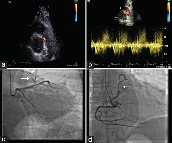
Congenital coronary-pulmonary fistulas (CPFs) are defined as an abnormal direct communication between one or more coronary arteries, with a cardiac or thoracic structure bypassing the capillary network. We report the case of a 73-year-old male, with a history of hypertension, asymptomatic for angina and dyspnea, who was referred for routine clinical control. Echocardiogram unexpectedly showed the presence of diastolic flow from the pulmonary trunk in parasternal short-axis view. Pulsed-wave Doppler confirmed the presence of diastolic flow and displayed the typical coronary flow pattern. Coronary angiography hence showed the presence of dual CPFs originating from the second segment of the left anterior descending coronary and the right coronary arteries. Careful routine Doppler echocardiograph examination may raise the suspicion of CPF in case of otherwise unexplained symptoms and may represent a simple, easy, repeatable tool for the first suspected diagnosis and follow-up of CPFs.
Keywords: Coronary anomaly; coronary fistula; doppler echocardiogram.
Conflict of interest statement
There are no conflicts of interest.
Figures
Similar articles
-
Left anterior descending coronary artery originating from the pulmonary artery: a rarity suspected during echocardiography.Turk Kardiyol Dern Ars. 2008 Apr;36(3):181-3. Turk Kardiyol Dern Ars. 2008. PMID: 18626212
-
The characteristics of coronary-pulmonary artery fistulas and the effectivity of trans-catheter closure: a single center experience.J Thorac Dis. 2019 Jul;11(7):2808-2815. doi: 10.21037/jtd.2019.06.60. J Thorac Dis. 2019. PMID: 31463109 Free PMC article.
-
Coronary artery-pulmonary artery fistula: case report.Cardiovasc Ultrasound. 2007 Apr 11;5:19. doi: 10.1186/1476-7120-5-19. Cardiovasc Ultrasound. 2007. PMID: 17428337 Free PMC article.
-
The role of the fractional flow reserve in the coronary steal phenomenon evaluation caused by the coronary-pulmonary fistulas: case report and review of the literature.J Cardiothorac Surg. 2020 Feb 3;15(1):32. doi: 10.1186/s13019-020-1073-x. J Cardiothorac Surg. 2020. PMID: 32013986 Free PMC article. Review.
-
Coronary artery fistulas: clinical and therapeutic considerations.Int J Cardiol. 2006 Feb 8;107(1):7-10. doi: 10.1016/j.ijcard.2005.01.067. Epub 2005 Aug 24. Int J Cardiol. 2006. PMID: 16125261 Review.
References
-
- Sherwood MC, Rockenmacher S, Colan SD, Geva T. Prognostic significance of clinically silent coronary artery fistulas. Am J Cardiol. 1999;83:407–11. - PubMed
-
- Hsieh KS, Huang TC, Lee CL. Coronary artery fistulas in neonates, infants, and children: Clinical findings and outcome. Pediatr Cardiol. 2002;23:415–9. - PubMed
-
- Gillebert C, Van Hoof R, Van de Werf F, Piessens J, De Geest H. Coronary artery fistulas in an adult population. Eur Heart J. 1986;7:437–43. - PubMed