Potential Toxic Effect of Bisphenol A on the Cardiac Muscle of Adult Rat and the Possible Protective Effect of Omega-3: A Histological and Immunohistochemical Study
- PMID: 31008050
- PMCID: PMC6442328
- DOI: 10.4103/JMAU.JMAU_53_18
Potential Toxic Effect of Bisphenol A on the Cardiac Muscle of Adult Rat and the Possible Protective Effect of Omega-3: A Histological and Immunohistochemical Study
Abstract
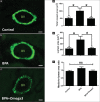
Bisphenol A (BPA) is intensely used in the production of polycarbonate plastics and epoxy resins. Recently, BPA has been receiving increased attention due to its link to various health problems that develop after direct or indirect human exposure. Previous studies have shown the harmful effect of high doses of BPA; however, the effect of small doses of BPA on disease development is controversial. The aim of this study was to investigate the effect of a low dose of BPA on the rat myocardium and to explore the outcome of coadministration of Omega-3 fatty acid (FA). Thirty adult male rats were divided equally into control group, BPA-treated group (1.2 mg/kg/day, intraperitoneally for 3 weeks), and BPA and Omega-3-treated group (received BPA as before plus Omega-3 at a daily dose of 300 mg/kg/day orally) for 3 weeks. Exposure to BPA resulted in structural anomalies in the rat myocardium in the form of disarrangement of myofibers, hypertrophy of myocytes, myocardial fibrosis, and dilatation of intramyocardial arterioles. On the other hand, mast cell density and media-to-lumen area ratio were not significantly altered. Interestingly, concomitant administration of Omega-3 FAs with BPA significantly reduced BPA-induced changes and provided a protective effect to the myocardium. In conclusion, exposure to a low dose of BPA could potentially lead to pathological alterations in the myocardium, which could be prevented by administration of Omega-3 FA.
Keywords: Bisphenol A; Omega-3; histopathology; myocardium.
Conflict of interest statement
There are no conflicts of interest.
Figures


Similar articles
-
Potential Effects of Bisphenol A on the Heart and Coronary Artery of Adult Male Rats and the Possible Role of L-Carnitine.J Toxicol. 2022 Dec 26;2022:7760594. doi: 10.1155/2022/7760594. eCollection 2022. J Toxicol. 2022. PMID: 36601412 Free PMC article.
-
Dose-dependent effect of Bisphenol-A on insulin signaling molecules in cardiac muscle of adult male rat.Chem Biol Interact. 2017 Mar 25;266:10-16. doi: 10.1016/j.cbi.2017.01.022. Epub 2017 Jan 30. Chem Biol Interact. 2017. PMID: 28153594
-
Low-dose developmental exposure to bisphenol A induces sex-specific effects in bone of Fischer 344 rat offspring.Environ Res. 2017 Nov;159:61-68. doi: 10.1016/j.envres.2017.07.020. Epub 2017 Aug 1. Environ Res. 2017. PMID: 28772150
-
Human exposure to bisphenol A.Toxicology. 2006 Sep 21;226(2-3):79-89. doi: 10.1016/j.tox.2006.06.009. Epub 2006 Jun 16. Toxicology. 2006. PMID: 16860916 Review.
-
Nephrotoxicity of bisphenol A (BPA)--an updated review.Curr Mol Pharmacol. 2013 Nov;6(3):163-72. doi: 10.2174/1874467207666140410115823. Curr Mol Pharmacol. 2013. PMID: 24720537 Review.
Cited by
-
Bisphenol A (BPA) Leading to Obesity and Cardiovascular Complications: A Compilation of Current In Vivo Study.Int J Mol Sci. 2022 Mar 9;23(6):2969. doi: 10.3390/ijms23062969. Int J Mol Sci. 2022. PMID: 35328389 Free PMC article. Review.
-
Cardiovascular and renal oxidative stress-mediated toxicities associated with bisphenol-A exposures are mitigated by Curcuma longa in rats.Avicenna J Phytomed. 2024 Mar-Apr;14(2):202-214. doi: 10.22038/AJP.2023.23367. Avicenna J Phytomed. 2024. PMID: 38966628 Free PMC article.
-
Curcumin ameliorated low dose-Bisphenol A induced gastric toxicity in adult albino rats.Sci Rep. 2022 Jun 17;12(1):10201. doi: 10.1038/s41598-022-14158-1. Sci Rep. 2022. PMID: 35715475 Free PMC article.
-
Potential Effects of Bisphenol A on the Heart and Coronary Artery of Adult Male Rats and the Possible Role of L-Carnitine.J Toxicol. 2022 Dec 26;2022:7760594. doi: 10.1155/2022/7760594. eCollection 2022. J Toxicol. 2022. PMID: 36601412 Free PMC article.
References
-
- Pivnenko K, Pedersen GA, Eriksson E, Astrup TF. Bisphenol A and its structural analogues in household waste paper. Waste Manag. 2015;44:39–47. - PubMed
-
- Welshons WV, Nagel SC, vom Saal FS. Large effects from small exposures. III. Endocrine mechanisms mediating effects of bisphenol A at levels of human exposure. Endocrinology. 2006;147:S56–69. - PubMed
-
- Vandenberg LN, Chahoud I, Heindel JJ, Padmanabhan V, Paumgartten FJ, Schoenfelder G. Urinary, circulating, and tissue biomonitoring studies indicate widespread exposure to bisphenol A. Cienc Saude Colet. 2012;17:407–34. - PubMed
-
- Teeguarden JG, Waechter JM, Jr, Clewell HJ, 3rd, Covington TR, Barton HA. Evaluation of oral and intravenous route pharmacokinetics, plasma protein binding, and uterine tissue dose metrics of bisphenol A: A physiologically based pharmacokinetic approach. Toxicol Sci. 2005;85:823–38. - PubMed
LinkOut - more resources
Full Text Sources
Research Materials
Miscellaneous
