

Optimal cut-off criteria for duplex ultrasound compared with computed tomography angiography for the diagnosis of restenosis in stented carotid arteries in the international carotid stenting study
- PMID: 31008301
- PMCID: PMC6453175
- DOI: 10.1177/2396987316678361
Optimal cut-off criteria for duplex ultrasound compared with computed tomography angiography for the diagnosis of restenosis in stented carotid arteries in the international carotid stenting study
Abstract
Introduction: Previous studies that reported duplex-ultrasound cut-off criteria, based on blood velocity parameters, for the degree of stenosis in a stented carotid artery were either retrospective, or the reference test was carried out only when a patient was suspected of having restenosis at duplex ultrasound, which is likely to have resulted in verification bias. We performed a prospective study of diagnostic accuracy to find new blood velocity cut-offs in duplex ultrasound for in-stent restenosis.
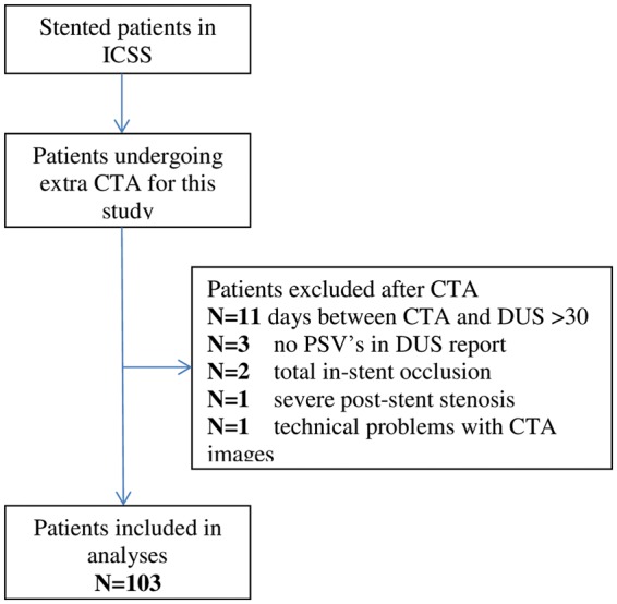
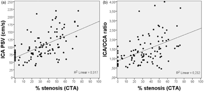
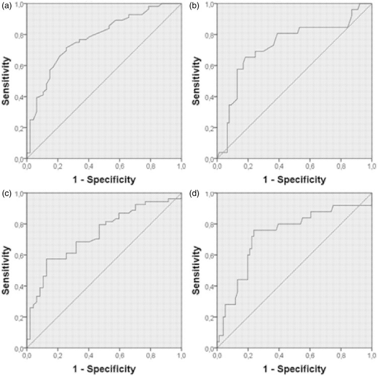
Patients and methods: Stented patients within the international carotid stenting study were eligible. Patients had a carotid computed tomography angiography in addition to routine duplex ultrasound performed at a yearly follow-up. Duplex-ultrasound bloodflow velocity parameters were compared to the degree of stenosis on computed tomography angiography. The results were analysed using receiver-operating-characteristic curves.
Results: We included 103 patients in this study. On computed tomography angiography, 30 (29.1%) patients had a 30%-49% in-stent restenosis, 21 (20.4%) patients had 50%-69% in-stent restenosis and 5 (4.9%) patients a ≥70% in-stent restenosis. The cut-off value ≥50% stenosis was a peak systolic velocity of 125 cm/s (sensitivity: 63% (95% CI: 41-79), specificity: 83% (95% CI: 72-90)).
Discussion: This study provides a level 2b evidence for new cut-off values for in-stent restenosis. Unfortunately, we could not say anything about severe stenosis because of the low number of severe stenosis after one year.
Conclusions: The 125 cm/s cut-off value on duplex ultrasound is lower than found in previous studies and equal to unstented arteries. Duplex-ultrasound measurements made in stented carotid arteries should not be corrected for the presence of a stent when determining the degree of stenosis.
Keywords: Stroke; computed tomography angiography; duplex ultrasound; restenosis; stenting.
Conflict of interest statement
The author(s) declared no potential conflicts of interest with respect to the research, authorship, and/or publication of this article.
Figures
References
-
- Hobson RW. Update on the Carotid Revascularization Endarterectomy vs. Stent Trial (CREST) protocol. J Am Coll Surg 2002; 194: S9–S14. - PubMed
-
- Featherstone RL, Brown MM, Coward LJ. ICSS Investigators. International carotid stenting study: protocol for a randomised clinical trial comparing carotid stenting with endarterectomy in symptomatic carotid artery stenosis. Cerebrovasc Dis 2004; 18: 69–74. - PubMed
-
- Wardlaw JM, Chappell FM, Best JJ, et al. NHS Research and Development Health Technology Assessment Carotid Stenosis Imaging Group. Non-invasive imaging compared with intra-arterial angiography in the diagnosis of symptomatic carotid stenosis: a meta-analysis. Lancet 2006; 367: 1503–1512. - PubMed
Grants and funding
LinkOut - more resources
Full Text Sources
