

Biplanar ultrasound investigation of in vivo Achilles tendon displacement non-uniformity
- PMID: 31008448
- PMCID: PMC6472705
- DOI: 10.1002/tsm2.61
Biplanar ultrasound investigation of in vivo Achilles tendon displacement non-uniformity
Abstract
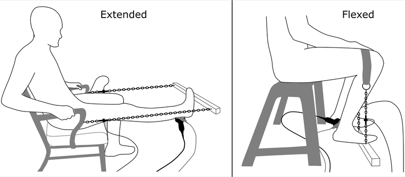
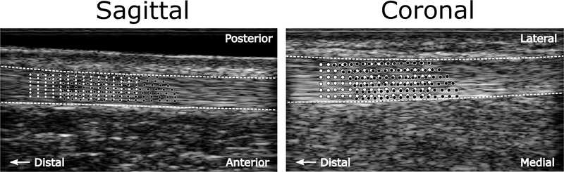
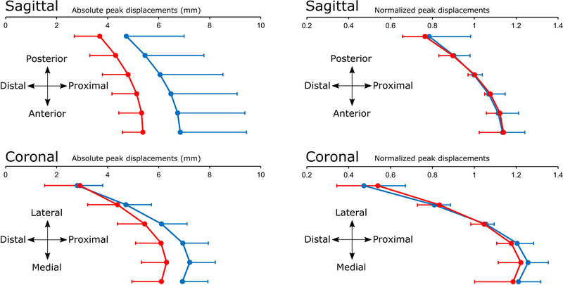
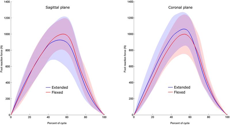
The Achilles tendon is a common tendon for the medial and lateral gastrocnemius and soleus muscles. Non-uniform Achilles tendon regional displacements have been observed in vivo which may result from non-uniform muscle loading and intra-tendinous shearing. However, prior observations are limited to the sagittal plane. This study investigated Achilles tendon tissue displacement patterns during isometric plantarflexor contractions in the coronal and sagittal planes. Fourteen subjects (5 female, 9 male, 26±3 yr) performed maximal isometric plantarflexor contractions with the knee in full extension and flexed to 110°. An ultrasound transducer positioned over the free Achilles tendon collected beam formed radio frequency (RF) data at 70 frames/s. Localized tissue displacements were analyzed using a speckle tracking algorithm. We observed non-uniform Achilles tendon tissue displacements in both imaging planes. Knee joint posture had no significant effect on tissue displacement patterns in either imaging plane. The non-uniform Achilles tendon tissue displacements during loading may arise from the anatomical organization of the sub-tendons associated with the three heads of the triceps surae. The biplanar investigation suggests that greatest displacements are localized to tissue likely to belong to soleus sub-tendon. This study adds novel information with possible implications for muscle coordination, function and muscle-tendon injury mechanisms.
Keywords: speckle tracking; sub-tendon; triceps surae; ultrasound.
Figures
Similar articles
-
Non-uniform displacements within the Achilles tendon observed during passive and eccentric loading.J Biomech. 2014 Sep 22;47(12):2831-5. doi: 10.1016/j.jbiomech.2014.07.032. Epub 2014 Aug 8. J Biomech. 2014. PMID: 25150898 Free PMC article.
-
Do triceps surae muscle dynamics govern non-uniform Achilles tendon deformations?PeerJ. 2018 Jul 12;6:e5182. doi: 10.7717/peerj.5182. eCollection 2018. PeerJ. 2018. PMID: 30013844 Free PMC article.
-
The effects of triceps surae muscle stimulation on localized Achilles subtendon tissue displacements.J Exp Biol. 2021 Aug 1;224(15):jeb242135. doi: 10.1242/jeb.242135. Epub 2021 Aug 5. J Exp Biol. 2021. PMID: 34350951 Free PMC article.
-
Triceps surae muscle-subtendon interaction differs between young and older adults.Connect Tissue Res. 2020 Jan;61(1):104-113. doi: 10.1080/03008207.2019.1612384. Epub 2019 May 22. Connect Tissue Res. 2020. PMID: 31116576 Free PMC article. Clinical Trial.
-
Local displacement within the Achilles tendon induced by electrical stimulation of the single gastrocnemius muscles.Clin Biomech (Bristol). 2023 Feb;102:105901. doi: 10.1016/j.clinbiomech.2023.105901. Epub 2023 Feb 2. Clin Biomech (Bristol). 2023. PMID: 36791484
Cited by
-
Individual variation in Achilles tendon morphology and geometry changes susceptibility to injury.Elife. 2021 Feb 16;10:e63204. doi: 10.7554/eLife.63204. Elife. 2021. PMID: 33588992 Free PMC article.
-
In vivo characterization of Achilles subtendon function and morphology within the tendon cross section and along the free tendon.bioRxiv [Preprint]. 2025 Jun 1:2025.05.29.656873. doi: 10.1101/2025.05.29.656873. bioRxiv. 2025. PMID: 40661625 Free PMC article. Preprint.
-
Achilles tendon and triceps surae muscle properties in athletes.Eur J Appl Physiol. 2024 Feb;124(2):633-647. doi: 10.1007/s00421-023-05348-4. Epub 2023 Nov 11. Eur J Appl Physiol. 2024. PMID: 37950761 Free PMC article.
-
Ultrasound speckle tracking of Achilles tendon in individuals with unilateral tendinopathy: a pilot study.Eur J Appl Physiol. 2020 Mar;120(3):579-589. doi: 10.1007/s00421-020-04317-5. Epub 2020 Feb 14. Eur J Appl Physiol. 2020. PMID: 32060739
-
Techniques for In Vivo Measurement of Ligament and Tendon Strain: A Review.Ann Biomed Eng. 2021 Jan;49(1):7-28. doi: 10.1007/s10439-020-02635-5. Epub 2020 Oct 6. Ann Biomed Eng. 2021. PMID: 33025317 Free PMC article. Review.
References
-
- Finni T, Komi PV, Lukkariniemi J. Achilles tendon loading during walking: application of a novel optic fiber technique. Eur J Appl Physiol Occup Physiol 1998;77:289–291. - PubMed
-
- Fukashiro S, Komi PV, Jarvinen M, Miyashita M. In vivo Achilles tendon loading during jumping in humans. Eur J Appl Physiol Occup Physiol 1995;71:453–458. - PubMed
-
- Kujala UM, Sarna S, Kaprio J. Cumulative incidence of achilles tendon rupture and tendinopathy in male former elite athletes. Clin J Sport Med 2005;15:133–135. - PubMed
Grants and funding
LinkOut - more resources
Full Text Sources