. 2019 May;20(5):731-747.
doi: 10.1111/mpp.12788.

A novel Botrytis cinerea-specific gene BcHBF1 enhances virulence of the grey mould fungus via promoting host penetration and invasive hyphal development

Affiliations

A novel Botrytis cinerea-specific gene BcHBF1 enhances virulence of the grey mould fungus via promoting host penetration and invasive hyphal development

Yue Liu et al. Mol Plant Pathol. 2019 May.

Abstract

Botrytis cinerea is the causative agent of grey mould on over 1000 plant species and annually causes enormous economic losses worldwide. However, the fungal factors that mediate pathogenesis of the pathogen remain largely unknown. Here, we demonstrate that a novel B. cinerea-specific pathogenicity-associated factor BcHBF1 (hyphal branching-related factor 1), identified from virulence-attenuated mutant M8008 from a B. cinerea T-DNA insertion mutant library, plays an important role in hyphal branching, infection structure formation, sclerotial formation and full virulence of the pathogen. Deletion of BcHBF1 in B. cinerea did not impair radial growth of mycelia, conidiation, conidial germination, osmotic- and oxidative-stress adaptation, as well as cell wall integrity of the ∆Bchbf1 mutant strains. However, loss of BcHBF1 impaired the capability of hyphal branching, appressorium and infection cushion formation, appressorium host penetration and virulence of the pathogen. Moreover, disruption of BcHBF1 altered conidial morphology and dramatically impaired sclerotial formation of the mutant strains. Complementation of BcHBF1 completely rescued all the phenotypic defects of the ∆Bchbf1 mutants. During young hyphal branching, host penetration and early invasive growth of the pathogen, BcHBF1 expression was up-regulated, suggesting that BcHBF1 is required for these processes. Our findings provide novel insights into the fungal factor mediating pathogenesis of the grey mould fungus via regulation of its infection structure formation, host penetration and invasive hyphal branching and growth.

Keywords: Botrytis cinerea; appressorium; host penetration; hyphal branching; infection cushions; sclerotial formation; virulence.


Figures

Figure 1
Figure 1
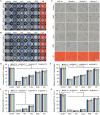
BcHBF1 is a pathogenicity‐associated gene. (A) Schematic diagram indicates the position of T‐DNA insertion in B. cinerea genome of the mutant M8008. (B) Expression of BcHBF1 in B. cinerea during conidial germination and the subsequent hyphal development. (C) Expression profile of B. cinerea BcHBF1 during host infection by the pathogen. (D) Strategy for generation of BcHBF1 gene disruption (∆Bchbf1) mutant strains. (E) Detection of BcHBF1 expression in the wild type (WT) (B05.10), ∆Bchbf1 and complemented (∆Bchbf1‐C) strains via real‐time quantitative Reverse Transcription‐Polymerase Chain Reaction (qRT‐PCR. (F) Pathogenicity assay for the WT, T‐DNA insertional mutant M8008, ∆Bchbf1 mutant and complemented strains. Droplets of conidial suspension (mixture of conidial suspension [1 × 106 conidia/mL] and PDB, vol: vol = 1: 1, 5 µL) of each strain were inoculated. Diseased leaves were photographically documented at 72 h post‐inoculation/incubation (hpi) at 20 °C in dark. (G) Quantification of lesion size caused by the indicated strains at 72 hpi. Data represent means ± standard deviations (SDs) from at least three independent experiments with triplicate samples examined for each treatment. ***: significant at P < 0.001.
Figure 2
Figure 2
BcHBF1 is required for B. cinerea conidial morphogenesis but dispensable for conidiation. (A) BcHBF1 mediates B. cinerea conidiation (upper panel) and conidial morphogenesis (lower panel). The indicated B. cinerea WT, ∆Bchbf1 and complemented strains were incubated on CM plates at 20 °C for 10 days and photographically documented. (B) Quantification of relative conidiation of the indicated strains at 10 days post‐incubation/inoculation (dpi) on CM plates. (C) Comparison of conidial size of the indicated strains. (D) Loss of BcHBF1 increases the number of globose and less elliptical conidia (closer to the dash line) in the ∆Bchbf1 mutant strains. More than 100 10‐day‐old conidia of each strain were measured under a microscope in each experiment. Data represent means ±  standard deviations (SDs) from three independent experiments with triplicate colonies/slides were analyzed for each strain. *** indicates significant at P < 0.001.
Figure 3
Figure 3
BcHBF1 is dispensable for B. cinerea conidial germination but required for hyphal branching. (A) Droplets of conidial suspension (5 × 105 conidia/mL, 10 µL) of each strain were inoculated on solid CM plates and incubated at 20 °C. Germinated conidia were photographically documented at the indicated hpi. (B and C) Quantification of hyphal branching (B) and total hyphal length (C) of the indicated strains at the indicated hpi. Data represent means ± standard deviations (SDs) from three independent experiments with triplicate plates examined for each treatment. ***: significant at P < 0.001.
Figure 4
Figure 4
Disruption of BcHBF1 in B. cinerea impairs sclerotial production. (A) Conidia of the wild type (WT), ∆Bchbf1, and ∆Bchbf1‐C strains were inoculated on CM plates at 20 °C in darkness. Sclerotial production by each strain was observed at the indicated dpi. (B) Quantification of sclerotial formation by the indicated strains during a time course of 30 days of incubation. ND: Not detected. (C) Quantification of the sizes of sclerotia produced by the indicated strains via ImageJ (https://imagej.nih.gov/ij/). (D) Germination of sclerotia produced by the indicated strains. The representative images are from one of the experiments, at least three independent experiments were performed, and all the experiments resulted in similar results. Data represent means ±  standard deviations (SDs) from three independent experiments in which triplicate plates were analyzed for each strain in each experiment. ***: significant at P < 0.001.
Figure 5
Figure 5
BcHBF1 is dispensable for osmotic‐ and oxidative‐stress adaptation as well as cell wall integrity of B. cinerea. (A) Conidial germination (1 × 106 conidia/mL, 1 µL) and hyphal development of the indicated wild type (WT), ∆Bchbf1, and ∆Bchbf1‐C strains of B. cinerea on CM plates supplemented with the osmotic stress agents NaCl (1 M) and KCl (1 M), the oxidative‐stress agent H2O2 (5 mM), and the cell wall disturbing agents sodium dodecyl sulfate (SDS, 0.005%) and Congo Red (CR, 300 μg/mL). (B) Mycelial radial growth (inoculated with fresh mycelial plugs, 5 mm in diameter) of the indicated strains on CM plates containing the indicated stress‐mimetic agents as presented in (A). Representative photographs were taken at 4 dpi. (C) Conidial germination (5 × 105 conidia/mL, 2 µL) of the indicated strains incubated on CM plates containing the assorted stress agents as presented in (A) for 6 h. (D and E) Quantification of the relative mycelial growth of the indicated strains growing from inoculated conidia (D) or mycelial plugs (5 mm in diameter) (E) on CM plates supplemented with the indicated stress‐mimetic agents as presented in (A). (F and G) Quantification of the relative conidial germination rates (F) and germ tube development (G) of the indicated strains incubated for 6 h on CM plates containing the indicated stress agents as presented in (A). Representative images are from one experiment, at least three independent experiments were performed and all the experiments resulted in similar results. Data represent means ± standard deviations (SDs) from three independent experiments in which triplicate plates were examined for each strain in each experiment. ***: significant at P < 0.001.
Figure 6
Figure 6
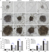
Loss of BcHBF1 reduces appressorium formation in B. cinerea. (A) BcHBF1 is required for B. cinerea appressorium formation and addition of fructose promotes the structure formation in the ∆Bchbf1 mutant strains. (B and C) Quantification of appressorium formation in the absence or presence of low concentration (1 mM) of fructose (B) and of higher concentration (10 mM) of fructose (C). For each strain, more than 100 conidia were examined in each experiment. Data represent means ± standard deviations (SDs) from three independent experiments. ***: significant at P < 0.001.
Figure 7
Figure 7
Loss of BcHBF1 impairs infection cushion development in B. cinerea. (A) Infection cushion formation of the indicated B. cinerea WT, ∆Bchbf1 and ∆Bchbf1‐C strains during a time course (36 h) of incubation at 20 ℃. Representative images are from one of three independent experiments; all the experiments resulted in similar results. (B and C) Quantification of the sizes (B) and numbers (C) of infection cushions produced by the indicated strains at the indicated hpi. Data represent means ± standard deviations (SDs) from three independent experiments in which triplicate slides were analyzed for each strain in each experiment. ***: significant at P < 0.001.
Figure 8
Figure 8
BcHBF1 is required for full virulence of B. cinerea. (A, B) Diseased green bean leaves caused by the indicated B. cinerea WT, ∆Bchbf1 and complemented strains during a time course (84 h) of infection. Droplets of conidial suspension (mixture of conidial suspension [1 × 106 conidia/mL] and PDB, vol: vol = 1: 1, 5 µL) (A) and mycelial plugs (5 mm in diameter) (B) of each strain were inoculated. The inoculated leaves were incubated at 20 °C in dark and diseased leaves were photographically documented at the indicated hpi. (C and D) Quantification of the lesion sizes caused by the indicated strains shown in (A) and (B), respectively. (E) Loss of BcHBF1 in B. cinerea impairs virulence of the pathogen on apple fruit via wound‐inoculation approach. (F) Quantification of the lesion sizes caused by the indicated strains shown in (E). Representative images are from one experiment. Data represent means ± standard deviations (SDs) from at least four independent experiments. *, ***: significant at P < 0.05, 0.001, respectively.
Figure 9
Figure 9
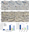
Loss of BcHBF1 impairs B. cinerea host penetration and invasive hyphal development. (A) Loss of BcHBF1 delays plant‐tissue penetration by the ∆Bchbf1 mutants. Droplets (10 µL) of conidial suspension (mixture of conidial suspension [1 × 105 conidia/mL] and ddH2O, vol: vol = 1: 10) of each strain were inoculated on onion epidermis (with extensive wash). The inoculated epidermis were incubated at 20 °C in dark; and at the indicated time points post‐inoculation, the inoculated epidermis were performed lactophenol blue staining and then photographically documented. (B) Disruption of BcHBF1 impairs B. cinerea invasive hyphal in planta branching and growth during infection. Bars in upper and lower panels are 40 µm and 100 µm, respectively. (C) Quantification of host penetration by the indicated strains at 12 hpi and 18 hpi. (D) Quantification of branch number of invasive hyphae of the indicated strains at 24 hpi. Representative images are from one of three independent experiments. Red arrows: appressoria and/or penetration points. Yellow arrows: branched or unbranched invasive hyphae. Co: conidium. IH: invasive hyphae. Data represent means ± standard deviations (SDs) from at least three independent experiments. ***: significant at P < 0.001.

Similar articles

Cited by

References

    1. Amselem, J. , Cuomo, C.A. , van Kan, J.A.L. , Viaud, M. , Benito, E.P. , Couloux, A. , Coutinho, P.M. , de Vries, R.P. , Dyer, P.S. , Fillinger, S. , Fournier, E. , Gout, L. , Hahn, M. , Kohn, L. , Lapalu, N. , Plummer, K.M. , Pradier, J.‐M. , Quévillon, E. , Sharon, A. , Simon, A. , ten Have, A. , Tudzynski, B. , Tudzynski, P. , Wincker, P. , Andrew, M. , Anthouard, V. , Beever, R.E. , Beffa, R. , Benoit, I. , Bouzid, O. , Brault, B. , Chen, Z. , Choquer, M. , Collémare, J. , Cotton, P. , Danchin, E.G. , Da Silva, C. , Gautier, A. , Giraud, C. , Giraud, T. , Gonzalez, C. , Grossetete, S. , Güldener, U. , Henrissat, B. , Howlett, B.J. , Kodira, C. , Kretschmer, M. , Lappartient, A. , Leroch, M. , Levis, C. , Mauceli, E. , Neuvéglise, C. , Oeser, B. , Pearson, M. , Poulain, J. , Poussereau, N. , Quesneville, H. , Rascle, C. , Schumacher, J. , Ségurens, B. , Sexton, A. , Silva, E. , Sirven, C. , Soanes, D.M. , Talbot, N.J. , Templeton, M. , Yandava, C. , Yarden, O. , Zeng, Q. , Rollins, J.A. , Lebrun, M.‐H. and Dickman, M . (2011) Genomic analysis of the necrotrophic fungal pathogens Sclerotinia sclerotiorum and Botrytis cinerea . PLoS Genet. 7, e1002230. - PMC - PubMed
    1. Cao, S.N. , Yuan, Y. , Qin, Y.H. , Zhang, M.Z. , de Figueiredo, P. , Li, G.H. and Qin, Q.M. (2018) The pre‐rRNA processing factor Nop53 regulates fungal development and pathogenesis via mediating production of reactive oxygen species. Environ. Microbiol. 20, 1531–1549. - PubMed
    1. Caracuel‐Rios, Z. and Talbot, N.J. (2007) Cellular differentiation and host invasion by the rice blast fungus Magnaporthe grisea . Curr. Opin. Microbiol. 10, 339–345. - PubMed
    1. Choquer, M. , Fournier, E. , Kunz, C. , Levis, C. , Pradier, J.‐M. , Simon, A. and Viaud, M. (2007) Botrytis cinerea virulence factors: new insights into a necrotrophic and polyphageous pathogen. FEMS Microbiol. Lett. 277, 1–10. - PubMed
    1. Dean, R. , Van Kan, J.A. , Pretorius, Z.A. , Hammond‐Kosack, K.E. , Di Pietro, A. , Spanu, P.D. , Rudd, J.J. , Dickman, M. , Kahmann, R. and Ellis, J. (2012) The Top 10 fungal pathogens in molecular plant pathology. Mol. Plant Pathol. 13, 414–430. - PMC - PubMed

Publication types

Substances

LinkOut - more resources