. 2019 May 14;58(19):2432-2446.
doi: 10.1021/acs.biochem.8b01272. Epub 2019 May 1.

Hydrogen-Deuterium Exchange Supports Independent Membrane-Interfacial Fusion Peptide and Transmembrane Domains in Subunit 2 of Influenza Virus Hemagglutinin Protein, a Structured and Aqueous-Protected Connection between the Fusion Peptide and Soluble Ectodomain, and the Importance of Membrane Apposition by the Trimer-of-Hairpins Structure

Affiliations

Hydrogen-Deuterium Exchange Supports Independent Membrane-Interfacial Fusion Peptide and Transmembrane Domains in Subunit 2 of Influenza Virus Hemagglutinin Protein, a Structured and Aqueous-Protected Connection between the Fusion Peptide and Soluble Ectodomain, and the Importance of Membrane Apposition by the Trimer-of-Hairpins Structure

Ahinsa Ranaweera et al. Biochemistry. .

Abstract

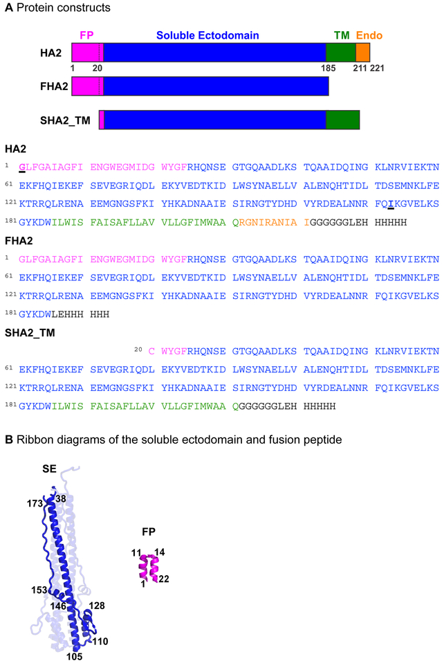
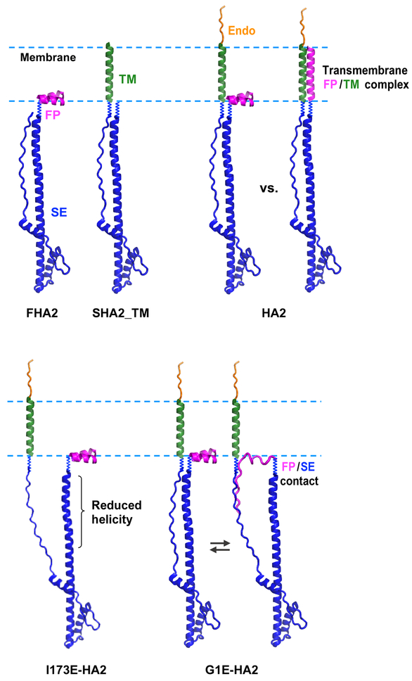
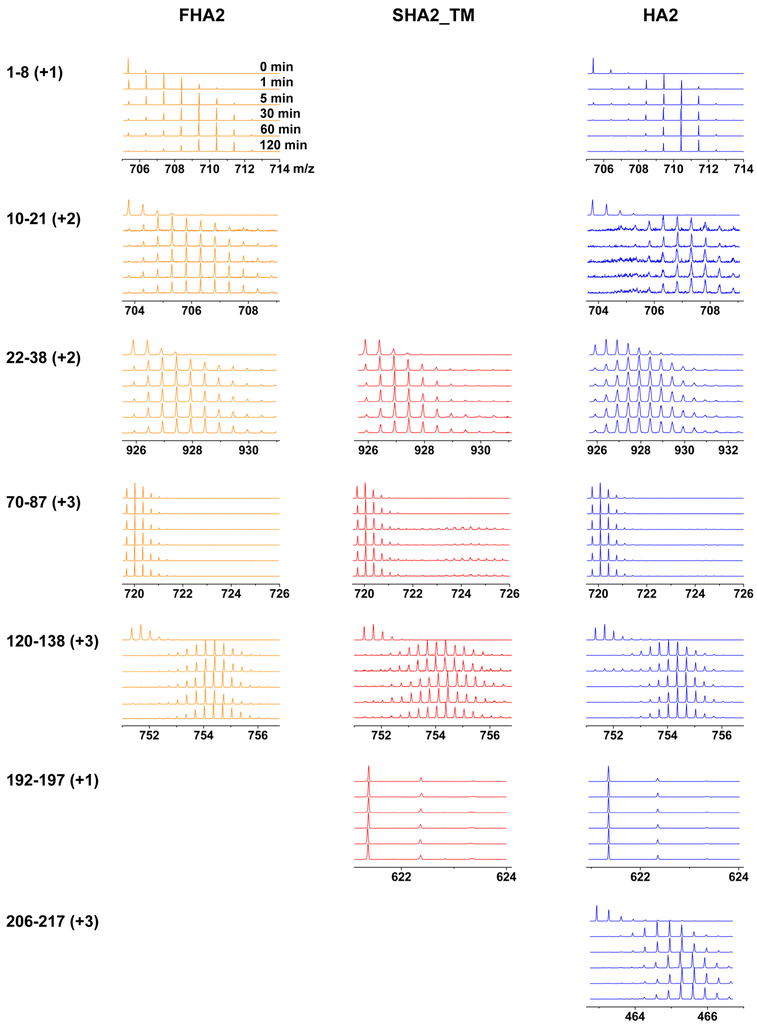
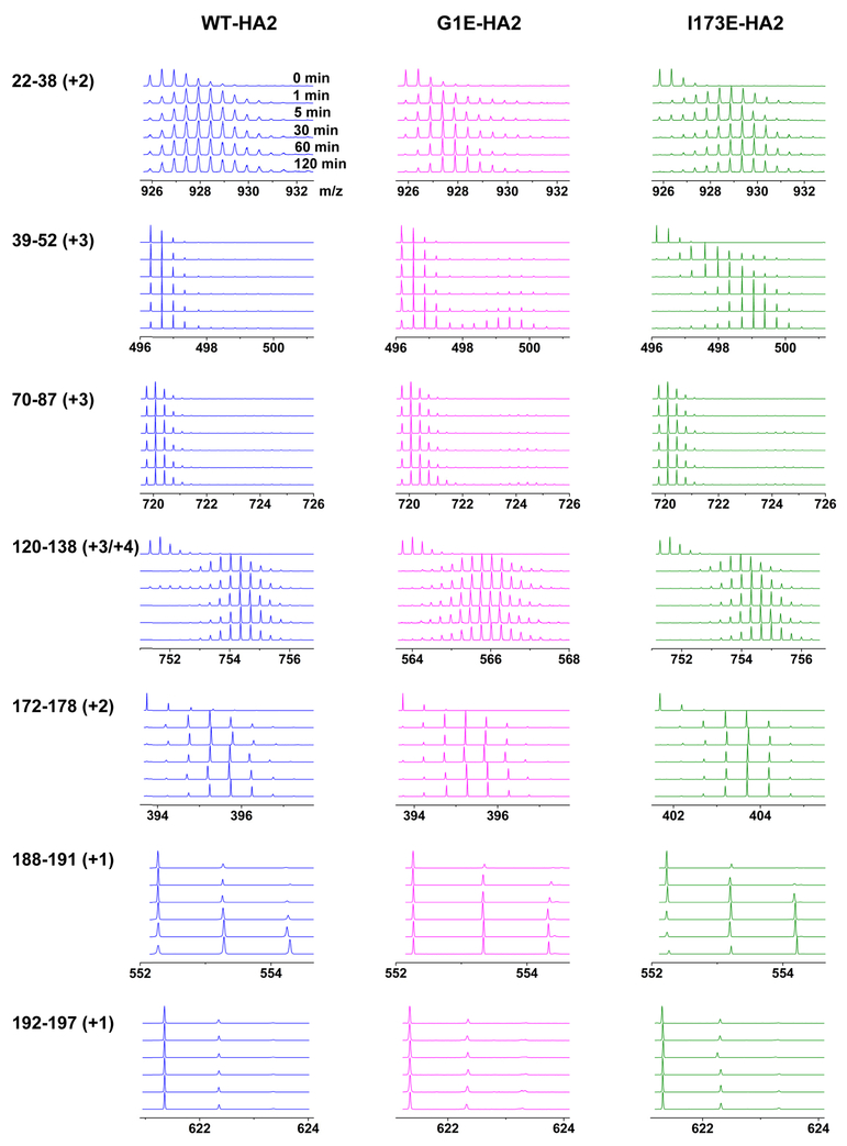
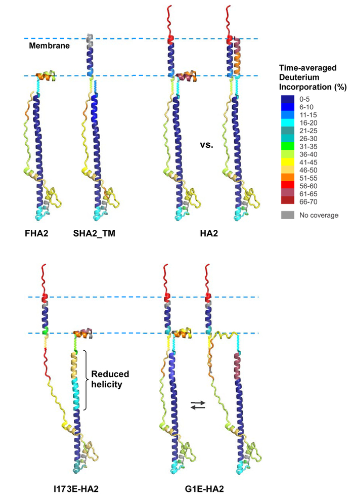
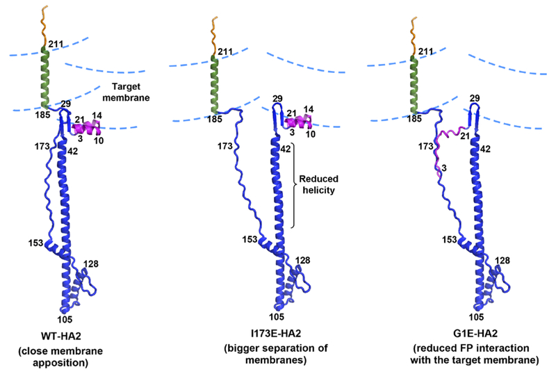
The influenza virus hemagglutinin (HA) protein has HA1 and HA2 subunits, which form an initial complex. HA1's bind host cell sialic acids, which triggers endocytosis, HA1/HA2 separation, and HA2-mediated fusion between virus and endosome membranes. We report hydrogen-deuterium exchange mass spectrometry (HDX-MS) on the HA2 subunit without HA1. HA2 contains the fusion peptide (FP), soluble ectodomain (SE), transmembrane domain (TM), and endodomain. FP is a monomer by itself, while SE is a trimer of hairpins that includes an interior bundle of residue 38-105 helices, turns, and residue 154-178 strands packed antiparallel to the bundle. FP and TM extend from the same side of the SE hairpin, and fusion models often depict a FP/TM complex with membrane traversal of both domains that is important for membrane pore expansion. The HDX-MS data of this study do not support this complex and instead support independent FP and TM with respective membrane-interfacial and traversal locations. The data also show a low level of aqueous exposure of the 22-38 segment, consistent with retention of the 23-35 antiparallel β sheet observed in the initial HA1/HA2 complex. We propose the β sheet as a semirigid connector between FP and SE that enables close membrane apposition prior to fusion. The I173E mutant exhibits greater exchange for residues 22-69 and 150-191, consistent with dissociation of SE C-terminal strands from interior N-helices. Similar trends are observed for the G1E mutant as well as less exchange for G1E FP. Fusion is highly impaired with either mutant, which correlates with reduced membrane apposition and, for G1E, FP binding to SE rather than the target membrane.


Figures

Figure 1.
Figure 1.
(A) Schematic diagrams and sequences of full-length HA2 and truncated constructs with domains colored: fusion peptide (FP), pink; soluble ectodomain (SE), blue; transmembrane domain (TM), green; and endodomain (Endo), orange. This color-coding is also used in Figures 2 and 8. Amino acid sequences are from the X31 strain which is a H3 subtype. The C-terminal regions in black include a non-native H6 tag for affinity chromatography. The G1E and I173E mutation sites are underlined in the HA2 sequence. (B) Ribbon diagrams of the SE in the final trimer-of-hairpins structure (PDB 1QU1) and the FP with closed helical hairpin structure (PDB 2KXA). A monomer subunit is highlighted for the SE, as well as the terminal residues of regular secondary structure elements, and residue 173.
Figure 2.
Figure 2.
Structural models for WT and mutant HA2 constructs that are based on residue 1–22 FP closed helical hairpin (PDB 2KXA), 36–175 SE hairpin (PDB 1QU1), 186–211 TM continuous α helix that traverses the membrane, and 212–221 endodomain that is disordered. Residues 23–35 and 176–185 are represented as squiggly lines, because there isn’t electron density for these residues in PDB 1QU1 for all or some of the monomers of the HA223–185 construct. The WT SE is the trimer-of-hairpins like in Fig. 1B, but a single monomer SE is displayed for clarity. Two WT-HA2 models are shown that either have a FP with helical hairpin structure at the membrane-interface, or FP with continuous α helical structure in complex with TM and with membrane traversal. I173E-HA2 is shown with dissociated C-terminal SE strands and reduced helicity in the N-terminal SE. This model is consistent with reduced overall helicity at ambient temperature and reduced Tm for I173E vs. WT. G1E-HA2 is shown as an equilibrium between the WT structure and the I173E structure with unfolded FP’s that bind the C-terminal SE strands. This model is based on similar helicity at ambient temperature and reduced Tm for G1E vs. WT.
Figure 3.
Figure 3.
HDX-MS data for selected peptides of FHA2, SHA2_TM, and HA2 proteins in DM detergent. Plots of mass spectral signal intensity vs. m/z are displayed for all tHDX ≡ the incubation time of the protein in buffer with D2O. The identity and charge of each peptide is given in the left column, with identities confirmed by MS/MS analysis. The shifts of the distributions to larger m/z with tHDX reflect increasing H→D substitution in the segment of the protein corresponding to the peptide.
Figure 4.
Figure 4.
Plots of percent deuterium incorporation (D%) vs. log10[tHDX/min] for all analyzed peptides of FHA2, SHA2_TM, and HA2. Peak intensities vs. m/z (Fig. 3) are inputs for the HX-Express software, which calculates peak area weighted-average m/z, denoted MH for dilution into H2O buffer, or MD(t) for dilution into D2O buffer for duration t ≡ tHDX. The D%(t) = 100 × [MD(t) – MH]/N, where N is the number of backbone amide hydrogens in the peptide. Each point in a plot is the average of triplicate measurements, with typical RMSD of 1%. Tables S1 list all D%(t) and associated replicate uncertainties.
Figure 5.
Figure 5.
HDX-MS data for selected peptides of WT-, G1E-, and I173E-HA2 proteins in DM detergent. Plots of mass spectral signal intensity vs. m/z are displayed for all tHDX ≡ the incubation time of the protein in buffer with D2O. The identity and charge of each peptide is given in the left column, with identities confirmed by MS/MS analysis. The shifts of the distributions to larger m/z with tHDX reflect increasing H→D substitution in the segment of the protein corresponding to the peptide.
Figure 6.
Figure 6.
Plots of percent deuterium incorporation (D%) vs. log10[HDX time/min] for all analyzed peptides of WT-, G1E-, and I173E-HA2. Peak intensities vs. m/z (Fig. 5) are inputs for the HX-Express software, which calculates peak area weighted-average m/z, denoted MH for dilution into H2O buffer, or MD(t) for dilution into D2O buffer for duration t ≡ tHDX. The D%(t) = 100 × [MD(t) – MH]/N, where N is the number of backbone amide hydrogens in the peptide. Each point in a plot is the average of triplicate measurements, with typical RMSD of 1%. Tables S2 list all D%(t) and associated replicate uncertainties.
Figure 7.
Figure 7.
Presentation of peptide 〈D%〉 from Tables 1 and 2 using color-coding and the structural models of Fig. 2. The colors for WT-HA2 are based on the 〈D%〉 of peptides 1–9, 10–21 22–38, 39–52, 53–69, 70–87, 86–91, 92–98, 99–110, 110–115, 120–138, 139–141, 142–150, 151–167, 168–171, 172–178, 179–187, 188–191, 192–197, 198–202, 200–205, and 206–217. The colors for other constructs are based on the 〈D%〉 of subsets of these peptides with a few substitutions that include 3–9 for G1E-HA2 and 1–8 and 12–22 for I173E-HA2.
Figure 8.
Figure 8.
Structural models for WT-, I173E-, and G1E-HA2 developed using the HDX-MS data of the present study. The proteins are bound to two membranes prior to fusion using color coding of domains like in Figs. 1 and 2. WT has trimer-of-hairpins structure for the SE as in Fig. 1B, but only one monomer is shown for clarity. The approximate positions of specific residues are indicated by the numbers. The WT model combines residue 1–22 closed FP helical hairpin (PDB 2KXA), 23–35 antiparallel β sheet of the HA21–175/HA1 complex (PDB 2HMG), 36–178 hairpin SE structure (PDB 1QU1), 186–211 continuous α helix for TM, and 212–221 unstructured endodomain. The 179–185 extended structure is found in one monomer in PDB 1QU1, but there isn’t electron density for these residues in the other two monomers. The TM traverses the virus membrane, the FP is interfacially-bound to the target membrane, and the membranes are held in close apposition by the fully-structured and semi-rigid 23–185 SE. The close apposition reduces the activation energy for fusion. The HDX-MS data support new features in Fig. 8 vs. 2 that include the 23–35 β sheet, 179–185 structure, and respective membrane interfacial and traversal locations of the FP and TM rather than the FP/TM complex in Fig. 2. The Fig. 8 structure is retained in the final fused membrane which has local positive curvature that accommodates the FP and TM locations (see Fig. S2) and may be important for fusion pore expansion. The I173E model shows dissociation of the C-terminal strands from the trimer-of-hairpins and reduced helicity for the N-helices, with resulting larger average distance between the membranes, and impaired fusion catalysis. The G1E model shows binding of the FP to the C-terminal strands, which results in more-distant membrane apposition and weaker FP binding to the target membrane, and consequent impaired fusion catalysis. The displayed G1E structure is in equilibrium with the WT structure, like in Fig. 2.

Similar articles

Cited by

References

    1. White JM, Delos SE, Brecher M, and Schornberg K (2008) Structures and mechanisms of viral membrane fusion proteins: Multiple variations on a common theme, Crit. Rev. Biochem. Mol. Biol 43, 189–219. - PMC - PubMed
    1. Harrison SC (2015) Viral membrane fusion, Virology 479, 498–507. - PMC - PubMed
    1. Boonstra S, Blijleven JS, Roos WH, Onck PR, van der Giessen E, and van Oijen AM (2018) Hemagglutinin-mediated membrane fusion: A biophysical perspective, Ann. Revs. Biophys 47, 153–173. - PubMed
    1. Nobusawa E, Aoyama T, Kato H, Suzuki Y, Tateno Y, and Nakajima K (1991) Comparison of complete amino acid sequences and receptor binding properties among 13 serotypes of hemagglutinins of influenza A viruses, Virology 182, 475–485. - PubMed
    1. Wilson IA, Skehel JJ, and Wiley DC (1981) Structure of the haemagglutinin membrane glycoprotein of influenza virus at 3 A resolution, Nature 289, 366–373. - PubMed

Publication types

MeSH terms