

Case Reports
doi: 10.1097/MD.0000000000014718.
Neuronavigator-guided ventriculoscopic approach for symptomatic xanthogranuloma of the choroid plexus in the lateral ventricle
Affiliations
- PMID: 31008920
- PMCID: PMC6494259
- DOI: 10.1097/MD.0000000000014718
Item in Clipboard
Case Reports
Neuronavigator-guided ventriculoscopic approach for symptomatic xanthogranuloma of the choroid plexus in the lateral ventricle
Medicine (Baltimore).
2019 Apr.
Abstract
Xanthogranuloma of choroid plexus is an extremely rare, benign, and mostly asymptomatic intracranial lesion. We report a case of symptomatic lateral ventricular xanthogranuloma resected via a neuronavigator-guided ventriculoscopic approach. Then we review recent English medical literature and notice that craniotomies have been the most popular treatment. But our choice of a ventriculoscopic approach possesses unique advantages such as minimized neural tissue damage, shortened operative time, less blood loss, and safer access to central structures over conventional open surgeries. Informed consent has been obtained from the patient and his immediate family regarding this case report.
Figures
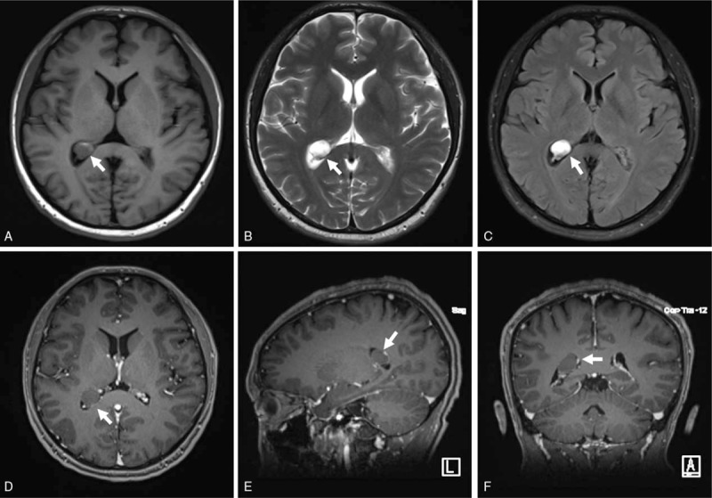
Preoperative magnetic resonance imaging showing a 1.3 × 1.2 × 1.0 cm oval mass filling the dilated posterior horn of the right lateral ventricle (white arrows). The lesion appeared hypointense on T1WI (A) including some hyperintense spots, and hyperintense on T2WI (B) as well as FLAIR images (C). Following contrast administration, no obvious enhancement was observed (D–F).

The patient's position, the scalp incision (A) and the burr hole (B). (C, D) A stereotactic navigation guidance system is used for a precise and straightforward trajectory to the lesion. (E) Structures of right lateral ventricle and the xanthogranuloma (XG) attached to the choroid plexus under endoscopic vision. (F) Choroid plexus after resection of XG in right lateral ventricle (black arrow).
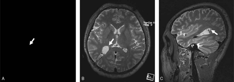
Postoperative magnetic resonance imaging showing no residual tumor (white arrows).

Photomicrographs of the surgical specimen. (A) Typical cholesterol clefts (arrows) and granulomas (arrowheads) enclosed by inflammatory cells (hematoxylin and eosin [H&E] stain, ×100). (B) Hyalinized and calcified blood vessels (asterisk) and psammoma bodies (arrowheads) (H&E stain, ×100). (C) Positive CD68 immunostaining in histiocytic cells surrounding cholesterol clefts and granulomas (H&E stain, ×100).
References
-
- Wolf A, Cowen D, Graham S. Xanthomas of the choroid plexus in man. J Neuropathol Exp Neurol 1950;9:286–97. - PubMed
-
- Giampalmo A, Viviano M. Cholesterosis of the choroid plexuses [undetermined language]. Accad Medica 1953;68:386–9. - PubMed
-
- Rosners S. Xanthoma of the choroid plexus in a child. J Nerv Ment Dis 1957;125:339–41. - PubMed
-
- Morello A, Campesi G, Bettinazzi N, et al. Neoplastiform xanthomatous granulomas of choroid plexus in a child affected by Hand-Schuller-Christian disease. Case report. J Neurosurg 1967;26:536–41. - PubMed
-
- Jaer O, Loken AC, Nesbakken R. Hydrocephalus due to xanthogranuloma. Case report. J Neurosurg 1973;39:659–61. - PubMed
Publication types
MeSH terms
LinkOut - more resources
Full Text Sources
Medical
