

Progressive sacroiliitis due to accessory sacroiliac joint mimicking ankylosing spondylitis: A case report
- PMID: 31008988
- PMCID: PMC6494240
- DOI: 10.1097/MD.0000000000015324
Progressive sacroiliitis due to accessory sacroiliac joint mimicking ankylosing spondylitis: A case report
Abstract
Rationale: An accessory sacroiliac (SI) joint usually has little clinical significance. However, severe arthritic changes can cause chronic buttock or low back pain and can be misdiagnosed as another disease presenting with sacroiliitis such as ankylosing spondylitis (AS).
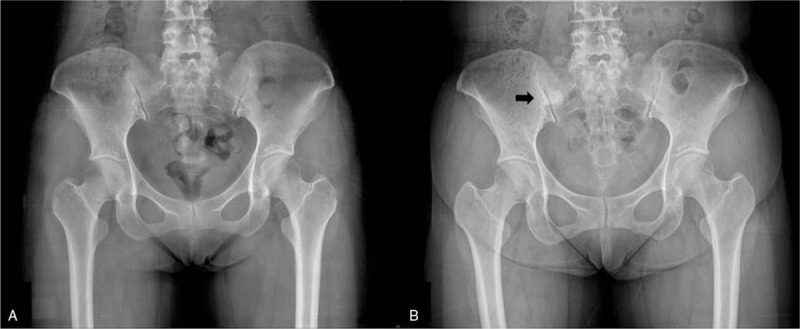
Patient concerns: A 33-year-old woman was diagnosed with AS due to chronic buttock pain and progressive sacroiliitis on plain X-ray and magnetic resonance imaging (MRI). Her buttock and low back pain gradually worsened despite proper treatment for AS.
Diagnosis: Computed tomography revealed an accessory SI joint with arthritic changes.
Interventions: Nonsteroidal anti-inflammatory drugs (NSAIDs) and restricted movement were prescribed.
Outcomes: The symptoms were controlled with NSAIDs, rest, and restriction of excessive movement. The medication could be stopped after the pain subsided.
Lessons: An accessory SI joint can be a cause of chronic back pain and can be misdiagnosed as AS with sacroiliitis when progressive arthritic changes are observed. Therefore, additional imaging studies other than conventional X-ray or MRI may be required for accurate diagnosis.
Conflict of interest statement
The authors have no conflicts of interest to disclose.
Figures


References
-
- Hadley LA. Accessory sacro-iliac articulations. J Bone Joint Surg Am 1952;34-A:149–55. - PubMed
-
- Rosa Neto NS, Vitule LF, Goncalves CR, et al. An accessory sacroiliac joint. Scand J Rheumatol 2009;38:496. - PubMed
-
- Prassopoulos PK, Faflia CP, Voloudaki AE, et al. Sacroiliac joints: anatomical variants on CT. J Comput Assist Tomogr 1999;23:323–7. - PubMed
-
- van der Linden S, Valkenburg HA, Cats A. Evaluation of diagnostic criteria for ankylosing spondylitis. A proposal for modification of the New York criteria. Arthritis Rheum 1984;27:361–8. - PubMed
-
- Rudwaleit M, van der Heijde D, Landewe R, et al. The development of Assessment of SpondyloArthritis international Society classification criteria for axial spondyloarthritis (part II): validation and final selection. Ann Rheum Dis 2009;68:777–83. - PubMed
Publication types
MeSH terms
LinkOut - more resources
Full Text Sources
Medical
Research Materials
