

Association Between Longitudinal Plasma Neurofilament Light and Neurodegeneration in Patients With Alzheimer Disease
- PMID: 31009028
- PMCID: PMC6583067
- DOI: 10.1001/jamaneurol.2019.0765
Association Between Longitudinal Plasma Neurofilament Light and Neurodegeneration in Patients With Alzheimer Disease
Erratum in
-
Error in Abstract.JAMA Neurol. 2019 Jul 1;76(7):872. doi: 10.1001/jamaneurol.2019.1649. JAMA Neurol. 2019. PMID: 31180462 Free PMC article. No abstract available.
Abstract
Importance: Plasma neurofilament light (NfL) has been suggested as a noninvasive biomarker to monitor neurodegeneration in Alzheimer disease (AD), but studies are lacking.
Objective: To examine whether longitudinal plasma NfL levels are associated with other hallmarks of AD.
Design, setting, and participants: This North American cohort study used data from 1583 individuals in the multicenter Alzheimer's Disease Neuroimaging Initiative study from September 7, 2005, through June 16, 2016. Patients were eligible for inclusion if they had NfL measurements. Annual plasma NfL samples were collected for up to 11 years and were analyzed in 2018.
Exposures: Clinical diagnosis, Aβ and tau cerebrospinal fluid (CSF) biomarkers, imaging measures (magnetic resonance imaging and fluorodeoxyglucose-positron emission tomography), and tests on cognitive scores.
Main outcomes and measures: The primary outcome was the association between baseline exposures (diagnosis, CSF biomarkers, imaging measures, and cognition) and longitudinal plasma NfL levels, analyzed by an ultrasensitive assay. The secondary outcomes were the associations between a multimodal classification scheme with Aβ, tau, and neurodegeneration (ie, the ATN system) and plasma NfL levels and between longitudinal changes in plasma NfL levels and changes in the other measures.
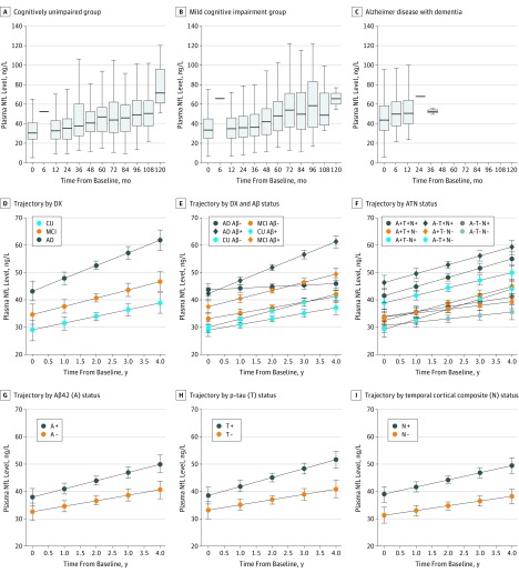
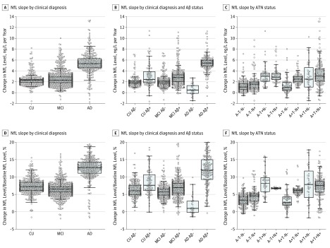
Results: Of the included 1583 participants, 716 (45.2%) were women, and the mean (SD) age was 72.9 (7.1) years; 401 had no cognitive impairment, 855 had mild cognitive impairment, and 327 had AD dementia. The NfL level was increased at baseline in patients with mild cognitive impairment and AD dementia (mean levels: cognitive unimpairment, 32.1 ng/L; mild cognitive impairment, 37.9 ng/L; and AD dementia, 45.9 ng/L; P < .001) and increased in all diagnostic groups, with the greatest increase in patients with AD dementia. A longitudinal increase in NfL level correlated with baseline CSF biomarkers (low Aβ42 [P = .001], high total tau [P = .02], and high phosphorylated tau levels [P = .02]), magnetic resonance imaging measures (small hippocampal volumes [P < .001], thin regional cortices [P = .009], and large ventricular volumes [P = .002]), low fluorodeoxyglucose-positron emission tomography uptake (P = .01), and poor cognitive performance (P < .001) for a global cognitive score. With use of the ATN system, increased baseline NfL levels were seen in A-T+N+ (P < .001), A+T-N+ (P < .001), and A+T+N+ (P < .001), and increased rates of NfL levels were seen in A-T+N- (P = .009), A-T+N+ (P = .02), A+T-N+ (P = .04), and A+T+N+ (P = .002). Faster increase in NfL levels correlated with faster increase in CSF biomarkers of neuronal injury, faster rates of atrophy and hypometabolism, and faster worsening in global cognition (all P < .05 in patients with mild cognitive impairment; associations differed slightly in cognitively unimpaired controls and patients with AD dementia).
Conclusions and relevance: The findings suggest that plasma NfL can be used as a noninvasive biomarker associated with neurodegeneration in patients with AD and may be useful to monitor effects in trials of disease-modifying drugs.
Conflict of interest statement
Figures
Comment in
-
The Neuroscientist Comments.Neuroscientist. 2019 Oct;25(5):385. doi: 10.1177/1073858419868688. Neuroscientist. 2019. PMID: 31554494 No abstract available.
References
Publication types
MeSH terms
Substances
Grants and funding
LinkOut - more resources
Full Text Sources
Other Literature Sources
Medical
Research Materials
Miscellaneous
