

Embryonic extracellular vesicles as informers to the immune cells at the maternal-fetal interface
- PMID: 31009068
- PMCID: PMC6718282
- DOI: 10.1111/cei.13304
Embryonic extracellular vesicles as informers to the immune cells at the maternal-fetal interface
Abstract
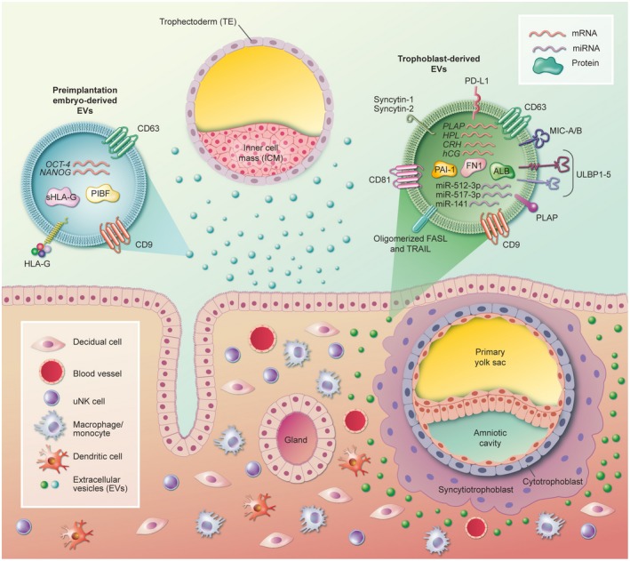
Extracellular vesicle (EV) exchange is emerging as a novel method of communication at the maternal-fetal interface. The presence of the EVs has been demonstrated in the preimplantation embryo culture medium from different species, such as bovines, porcines and humans. Preimplantation embryo-derived EVs have been shown to carry molecules potentially able to modulate the local endometrial immune system. The non-classical major histocompatibility complex (MHC) class I molecule human leucocyte antigen (HLA)-G, the immunomodulatory molecule progesterone-induced blocking factor and some regulatory miRNAs species are contained in embryo-derived EV cargo. The implanted syncytiotrophoblasts are also well known to secrete EVs, with microvesicles exerting a mainly proinflammatory effect while exosomes in general mediate local immunotolerance. This review focuses on the current knowledge on the potential role of EVs released by the embryo in the first weeks of pregnancy on the maternal immune cells. Collectively, the data warrant further exploration of the dialogue between the mother and the embryo via EVs.
Keywords: HLA-G; embryo; extracellular vesicles; maternal-fetal interface; trophoblast.
© 2019 British Society for Immunology.
Conflict of interest statement
The authors declare no conflicts of interest.
Figures
References
-
- Mathivanan S, Simpson RJ. ExoCarta: a compendium of exosomal proteins and RNA. Proteomics 2009; 9:4997–5000. - PubMed
Publication types
MeSH terms
LinkOut - more resources
Full Text Sources
Research Materials
