. 2019 Jun;46(6):2600-2609.
doi: 10.1002/mp.13554. Epub 2019 May 6.

Two-dimensional ultrasound-computed tomography image registration for monitoring percutaneous hepatic intervention

Affiliations

Two-dimensional ultrasound-computed tomography image registration for monitoring percutaneous hepatic intervention

Robert M Pohlman et al. Med Phys. 2019 Jun.

Abstract

Purpose: Deformable registration of ultrasound (US) and contrast enhanced computed tomography (CECT) images are essential for quantitative comparison of ablation boundaries and dimensions determined using these modalities. This comparison is essential as stiffness-based imaging using US has become popular and offers a nonionizing and cost-effective imaging modality for monitoring minimally invasive microwave ablation procedures. A sensible manual registration method is presented that performs the required CT-US image registration.

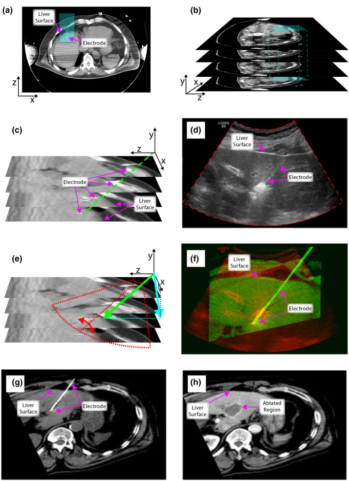
Methods: The two-dimensional (2D) virtual CT image plane that corresponds to the clinical US B-mode was obtained by "virtually slicing" the 3D CT volume along the plane containing non-anatomical landmarks, namely points along the microwave ablation antenna. The initial slice plane was generated using the vector acquired by rotating the normal vector of the transverse (i.e., xz) plane along the angle subtended by the antenna. This plane was then further rotated along the ablation antenna and shifted along with the direction of normal vector to obtain similar anatomical structures, such as the liver surface and vasculature that is visualized on both the CT virtual slice and US B-mode images on 20 patients. Finally, an affine transformation was estimated using anatomic and non-anatomic landmarks to account for distortion between the colocated CT virtual slice and US B-mode image resulting in a final registered CT virtual slice. Registration accuracy was measured by estimating the Euclidean distance between corresponding registered points on CT and US B-mode images.

Results: Mean and SD of the affine transformed registration error was 1.85 ± 2.14 (mm), computed from 20 coregistered data sets.

Conclusions: Our results demonstrate the ability to obtain 2D virtual CT slices that are registered to clinical US B-mode images. The use of both anatomical and non-anatomical landmarks result in accurate registration useful for validating ablative margins and comparison to electrode displacement elastography based images.

Keywords: computed tomography; microwave ablation monitoring; registration; ultrasound.


Conflict of interest statement

The authors have no conflicts to disclose.

Figures

Figure 1
Figure 1
Strategy for colocating and registering a computed tomography (CT) virtual slice from the 3D CT volume to the ultrasound B‐mode imaging plane.
Figure 2
Figure 2
Strategy for obtaining the computed tomography (CT) virtual slice from the 3D CT volume. Potential slice planes were obtained by varying a normal vector around antenna to acquire the plane corresponding to the ultrasound B‐mode image. [Color figure can be viewed at wileyonlinelibrary.com]
Figure 3
Figure 3
An example workflow using patient image data illustrating the ultrasound (US) B‐mode – computed tomography (CT) registration process. (a) Transverse slice with the antenna location highlighted in blue. (b) CT volume as a stack of transverse slices with the antenna location highlighted within the blue cube. (c) Zoomed in region of the antenna location in the CT volume with the antenna shown in green. (d) Comparable US B‐mode image from patient after ablation with outline of sector image highlighted in red. (e) Zoomed in CT volume with the two vectors creating the virtual slice plane highlighted in green and blue for the vector along the direction of the antenna and the vector projected onto the xy‐plane, respectively. An example sector US outline is transposed onto this plane which is rotated about the antenna to match three clearly visible anatomic landmarks. (f) The resultant CT virtual slice after affine transformation in green transposed onto the red US B‐mode image. (g) The entire CT virtual slice collected preablation. (h) The CT virtual slice estimated post‐ablation. [Color figure can be viewed at wileyonlinelibrary.com]
Figure 4
Figure 4
An example of ultrasound (US) B‐mode – computed tomography (CT) registration result for a second patient (a) US B‐mode prior to ablation with antenna visible. (b) Colocated CT virtual slice. (c) The registered CT virtual slice after affine transformation in green transposed onto the red. (d) The co‐located CT virtual slice estimated post‐ablation. [Color figure can be viewed at wileyonlinelibrary.com]
Figure 5
Figure 5
Computed tomography (CT) virtual slice registration accuracy to ultrasound (US) B‐mode. (a) Plot of non‐anatomical registration points (antenna) from US and CT. R2 = 0.98, P < 0.001. (b) Plot of anatomical registration points on US and CT. R2 = 0.98, P < 0.001. (c) Final plot of both anatomical and non‐anatomical points on CT and US. R2 = 0.98, P < 0.001. [Color figure can be viewed at wileyonlinelibrary.com]
Figure 6
Figure 6
Registration error distribution with translation, rotation, scaling rotation and translation (SRT) and affine transformation after the initial rigid registration (virtual slice) stage. Error is plotted on a log scale along the y‐axis. The ** denotes statistically significant improvement with affine transformation.

References

    1. Llovet JM, Fuster J, Bruix J and Barcelona‐Clinic Liver Cancer G . The Barcelona approach: diagnosis, staging, and treatment of hepatocellular carcinoma. Liver Transpl. 2004;10:S115–S120. - PubMed
    1. American Cancer Society . Cancer Facts and Figures. 2017;2017:2017.
    1. Okuda K, Liver Cancer Study Group of Japan . Primary liver cancers in Japan. Cancer. 1980;45:2663–2669. - PubMed
    1. Lai EC, Fan ST, Lo CM, Chu KM, Liu CL, Wong J. Hepatic resection for hepatocellular carcinoma. An audit of 343 patients. Ann Surg. 1995;221:291–298. - PMC - PubMed
    1. Potretzke TA, Ziemlewicz TJ, Hinshaw JL, et al. Microwave versus radiofrequency ablation treatment for hepatocellular carcinoma: a comparison of efficacy at a single center. J Vasc Interv Radiol. 2016;27:631–638. - PMC - PubMed

MeSH terms