. 2019 Apr 22;15(4):e1008093.
doi: 10.1371/journal.pgen.1008093. eCollection 2019 Apr.

Destabilization of chromosome structure by histone H3 lysine 27 methylation

Affiliations

Destabilization of chromosome structure by histone H3 lysine 27 methylation

Mareike Möller et al. PLoS Genet. .

Abstract

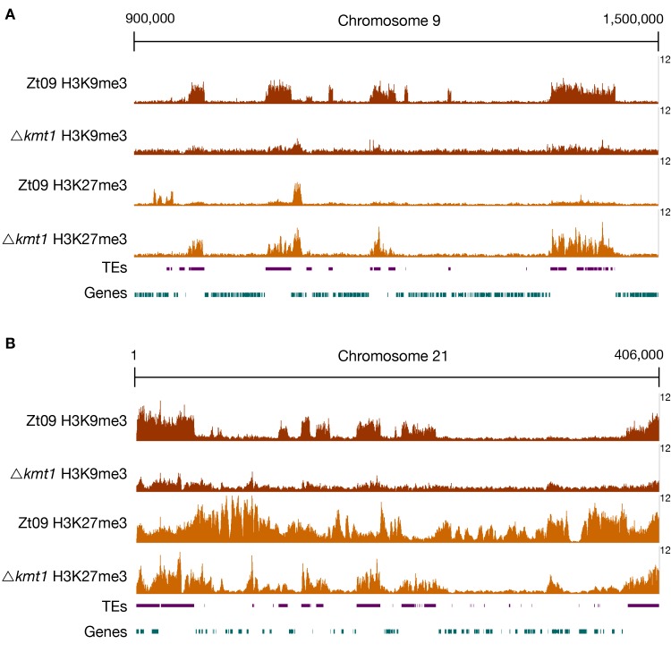
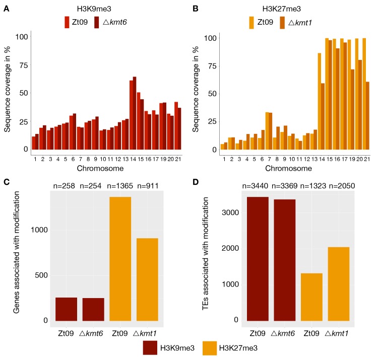
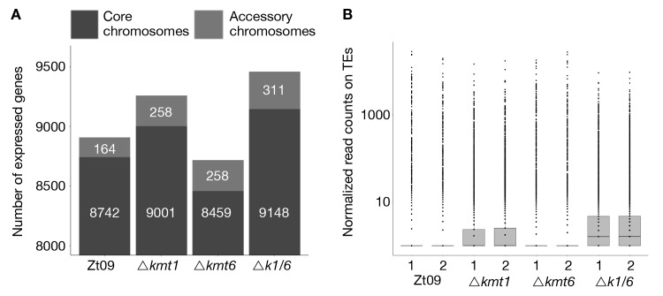
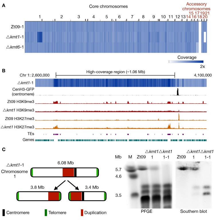
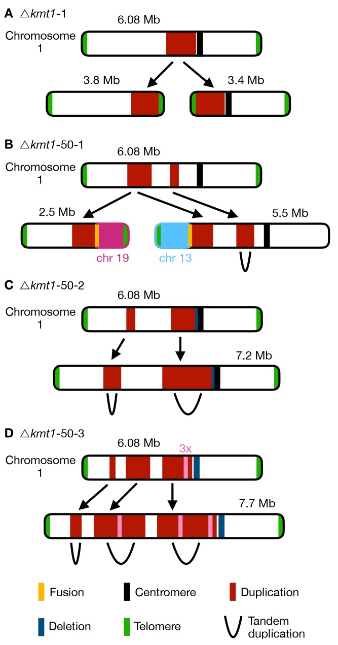
Chromosome and genome stability are important for normal cell function as instability often correlates with disease and dysfunction of DNA repair mechanisms. Many organisms maintain supernumerary or accessory chromosomes that deviate from standard chromosomes. The pathogenic fungus Zymoseptoria tritici has as many as eight accessory chromosomes, which are highly unstable during meiosis and mitosis, transcriptionally repressed, show enrichment of repetitive elements, and enrichment with heterochromatic histone methylation marks, e.g., trimethylation of H3 lysine 9 or lysine 27 (H3K9me3, H3K27me3). To elucidate the role of heterochromatin on genome stability in Z. tritici, we deleted the genes encoding the methyltransferases responsible for H3K9me3 and H3K27me3, kmt1 and kmt6, respectively, and generated a double mutant. We combined experimental evolution and genomic analyses to determine the impact of these deletions on chromosome and genome stability, both in vitro and in planta. We used whole genome sequencing, ChIP-seq, and RNA-seq to compare changes in genome and chromatin structure, and differences in gene expression between mutant and wildtype strains. Analyses of genome and ChIP-seq data in H3K9me3-deficient strains revealed dramatic chromatin reorganization, where H3K27me3 is mostly relocalized into regions that are enriched with H3K9me3 in wild type. Many genome rearrangements and formation of new chromosomes were found in the absence of H3K9me3, accompanied by activation of transposable elements. In stark contrast, loss of H3K27me3 actually increased the stability of accessory chromosomes under normal growth conditions in vitro, even without large scale changes in gene activity. We conclude that H3K9me3 is important for the maintenance of genome stability because it disallows H3K27me3 in regions considered constitutive heterochromatin. In this system, H3K27me3 reduces the overall stability of accessory chromosomes, generating a "metastable" state for these quasi-essential regions of the genome.


Conflict of interest statement

The authors have declared that no competing interests exist.

Figures

Fig 1
Fig 1
ChIP-seq reveals relocation of H3K27me3 on core (A) and accessory (B) chromosomes in Δkmt1 mutants. By analyzing ChIP-seq data in the Δkmt1 mutants we found that enrichment of H3K27me3 moves to sequences that are normally enriched with H3K9me3. A region on core chromosome 9 (A) is shown, where H3K27me3 is strongly enriched at former H3K9me3 regions, but depleted from its original positions. On accessory chromosomes (B), here full-length chromosome 21 as an example, there are similar dynamics as observed on core chromosomes. Accessory chromosomes normally show overall enrichment of H3K27me3. In absence of H3K9me3, H3K27me3 concentrates on former H3K9me3 regions, again being depleted from its original position. However, this effect varies between accessory chromosomes (Fig 2). The low amount of background found in Δkmt1 is due to the repetitive nature of the H3K9me3-enriched regions. All shown ChIP-seq tracks are normalized to 1x coverage (coverage indicated on the right) [107].
Fig 2
Fig 2. Genome-wide distribution of H3K9me3 and H3K27me3 in Zt09 and mutants.
(A) and (B) display the percentage of sequence coverage of core and accessory chromosomes with H3K9me3 and H3K27me3 relative to the chromosome length. While there were little differences in the overall coverage with H3K9me3 between Zt09 and Δkmt6 (A), H3K27me3 enrichment was increased on core chromosomes and decreased on accessory chromosomes in the Δkmt1 mutant (B). Chromosome 7 displayed a higher H3K27me3 coverage compared to the other core chromosomes as the right arm showed characteristics of an accessory chromosome [31]. (C and D) Genes (C) and TEs (D) associated with H3K9me3 or H3K27me3 in Zt09 and mutant strains. While there was almost no difference in terms of H3K9me3-associated genes or TEs in the Δkmt6 mutants, H3K27me3 relocated from genes to TEs in the Δkmt1 mutants.
Fig 3
Fig 3
Gene (A) and transposon (B) expression increases in absence of H3K9me3, while loss of H3K27me3 alone decreases the number of expressed genes and does not impact transposon activity. (A) We compared the number of expressed genes in Zt09 and mutant strains. While in all mutants the number of expressed genes increased on accessory chromosomes, surprisingly loss of H3K27me3 alone in the Δkmt6 mutants resulted in a reduction of genes expressed on core chromosomes and only a small increase in numbers of genes expressed on accessory chromosomes. (B) Loss of H3K9me3, but not H3K27me3 alone, increased the number of transcripts originating from TEs. In absence of both marks (Δk1/k6), the number further increased, likely because H3K27me3 moves to TEs in the Δkmt1 single mutant, facilitating silencing.
Fig 4
Fig 4. Genome sequencing of progenitor strains for the long-term growth experiment and analysis of structural variation in the Δkmt1 progenitor.
(A) The genomes of progenitor strains were sequenced and reads were mapped to the reference genome. Genome coverage was normalized to 1x coverage to allow identification and comparison of differences within and between strains. All strains were missing chromosome 18, as expected [41]. Δkmt6 had lower coverage (0.4x) of chromosome 17. Δkmt1 lost chromosome 20 and, most notably, showed a long segment (~ 1Mb) of high-coverage (1.6x) on chromosome 1. Centromeres are indicated as black dots. (B) Examination of the high-coverage region breakpoints on chromosome 1. The first breakpoint located within a TE-rich region that is enriched with H3K9me3 in Zt09 and showed new enrichment with H3K27me3 in Δkmt1. The second breakpoint is within a gene-rich region in close proximity to relocalized H3K27me3 and very close to the centromere (~15 kb). (C) Further analysis of this high-coverage region revealed de novo telomere formation at the breakpoints indicating a chromosome breakage at both ends of the high-coverage region. To validate chromosome breakage and possible new chromosome formation, we conducted PFGE and separated the large chromosomes of Zt09, of the Δkmt1 progenitor strain (Δkmt1-1) and of a single clone originating from the Δkmt1 progenitor strain stock (Δkmt1-1-1). Chromosome 1 (~6 Mb) is present in Zt09 and Δkmt1-1 (faint band), but not in the Δkmt1-1-1 single clone. We conducted Southern analysis on the PFGE blot using a sequence of the high-coverage region as a probe. It hybridized to the original chromosome 1 band in Zt09 and Δkmt1-1, but additionally to a ~3.4 Mb and ~3.8 Mb band in Δkmt1-1 and only to these bands in Δkmt1-1-1. This confirmed the formation of new chromosomes, both containing the high-coverage region in some cells of the progenitor strain population.
Fig 5
Fig 5. Genome sequencing of evolved populations and single clones originating from the long-term growth experiment.
(A) Genomes of each replicate population after 50 transfers were sequenced and mapped to the reference. Coverage is normalized to 1x. Except for coverage differences on the accessory chromosomes, there were no large structural variations detectable for the evolved Zt09 and Δkmt6 populations. In contrast, Δkmt1 populations contained multiple high-coverage regions (dark blue) on core chromosomes as well as large deletions indicated by low (light blue) or no (white) coverage. (B) To further characterize structural variation in the evolved Δkmt1 strains, three single clones originating from populations Δkmt1-50-1 (Δkmt1-50-1-1) and Δkmt1-50-2 (Δkmt1-50-2-1 and Δkmt1-50-2-2) were sequenced. Clones Δkmt1-50-1-1 and Δkmt1-50-2-2 show a very similar pattern as their respective populations, while Δkmt1-50-2-1 resembles a genotype that appears to be rare in population Δkmt1-50-2. High coverage on entire core chromosomes 13 (Δkmt1-50-2-1, 1.3x coverage) and 6 (Δkmt1-50-2-2, 1.5x coverage) indicates whole core chromosome duplications that were maintained in some nuclei. Centromeres are indicated as black dots.
Fig 6
Fig 6. Different outcomes of structural variation of chromosome 1 in evolved Δkmt1 strains.
Upper chromosome maps display coverage differences based on mapping to the reference genome including duplication indicated by higher read coverage (red) and deletion indicated by lower coverage (blue). The respective lower maps show structural rearrangements predicted by our structural variant analysis. (A) In the progenitor strain, a duplicated region was involved in the formation of two new chromosomes. At both termini of the duplication the chromosome broke and telomeric repeats were added de novo to these breakpoints (see Fig 4). Thus, two new chromosomes were formed, both containing the duplicated sequence. This structural variation was not found in all cells in the Δkmt1 progenitor strain and the structural variation that arose in the evolved strains (B-D) can therefore be the result of rearrangements of the reference chromosome 1 or the two newly formed chromosomes. (B) In the evolved population Δkmt1-50-1, two duplicated sequences were detected. The borders of the first region mark chromosome breakages that were fused to telomeres of other chromosomes. The first breakpoint was attached to the telomere of chromosome 13 forming a new 5.5 Mb chromosome while the second breakpoint was fused to the telomere of chromosome 19 (new 2.5 Mb chromosome). The second duplicated region represented a tandem duplication located on the new 5.5 Mb chromosome that falls within the duplicated region of the progenitor strain. (C) Population Δkmt1-50-2 contained two duplicated regions, that both resembled tandem duplications. The second duplication is very similar to the one found in the progenitor strain but includes half of the centromere and had a deletion, where the breakpoint close to the centromere in the progenitor strain is located. (D) Population Δkmt1-50-3 displayed three duplicated sequences that all form tandem duplications resulting in the formation of a 7.7 Mb version of chromosome 1. The third duplicated region was, as in population Δkmt1-50-2, very similar to the one in the progenitor strain. However, in this case the complete centromere-associated sequence was deleted. Furthermore, a ~50 kb region inside the third duplicated region exhibited 3x sequencing coverage and was found in between the tandem duplication of the second duplicated region (see S9 Table).
Fig 7
Fig 7. Working model to illustrate how H3K27me3 may influence nuclear localization of whole chromosomes or landmark genomic regions.
In wildtype cells (left panel), H3K27me3 is localized in subtelomeric regions and on accessory chromosomes, directing those regions to the nuclear periphery and resulting in increased instability of these regions. Loss of H3K27me3 (middle panel) results in a relocation of former H3K27me3-enriched sequences to the inner nucleus and an increase of genome stability. Loss of the histone modification H3K9me3 enables H3K27me3 to spread, leading to mislocalization of H3K27me3, altered physical localization and chromatin interactions in the nucleus that fuel genome instability of these regions (right panel).

Similar articles

Cited by

References

    1. Goldberg AD, Allis CD, Bernstein E. Epigenetics: A Landscape Takes Shape. Cell. 2007;128: 635–638. 10.1016/j.cell.2007.02.006 - DOI - PubMed
    1. Kornberg RD, Lorch Y. Chromatin structure and transcription. Annu Rev Cell Biol. 1992;8: 563–587. 10.1146/annurev.cb.08.110192.003023 - DOI - PubMed
    1. Venkatesh S, Workman JL. Histone exchange, chromatin structure and the regulation of transcription. Nat Rev Mol Cell Biol. 2015;16: 178–189. 10.1038/nrm3941 - DOI - PubMed
    1. Bannister AJ, Kouzarides T. Regulation of chromatin by histone modifications. Cell Res. 2011;21: 381–395. 10.1038/cr.2011.22 - DOI - PMC - PubMed
    1. Harr JC, Gonzalez‐Sandoval A, Gasser SM. Histones and histone modifications in perinuclear chromatin anchoring: from yeast to man. EMBO Rep. 2016;17: e201541809. - PMC - PubMed

Publication types