. 2019 Apr 22;15(4):e1007713.
doi: 10.1371/journal.ppat.1007713. eCollection 2019 Apr.

Clumping factor B is an important virulence factor during Staphylococcus aureus skin infection and a promising vaccine target

Affiliations

Clumping factor B is an important virulence factor during Staphylococcus aureus skin infection and a promising vaccine target

Keenan A Lacey et al. PLoS Pathog. .

Abstract

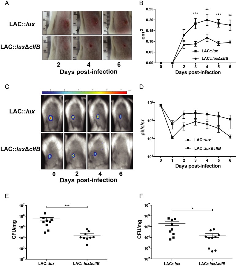
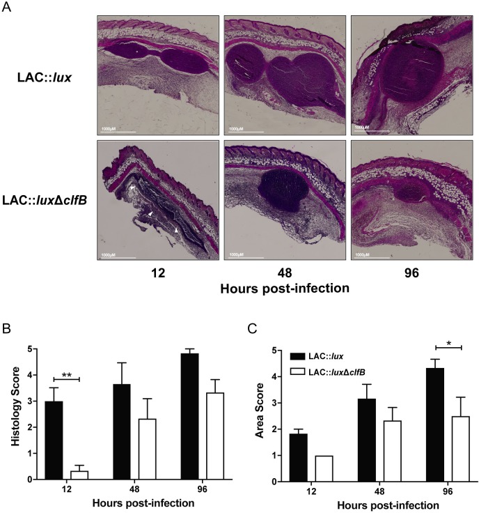
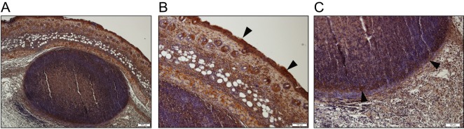
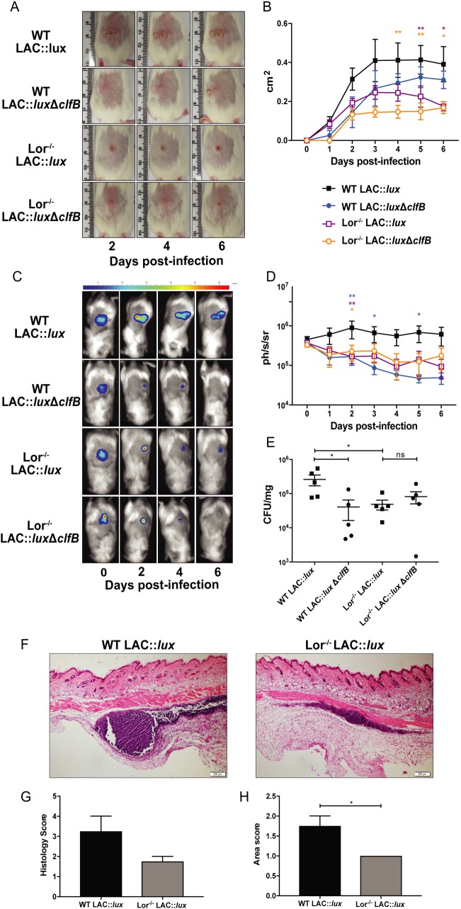
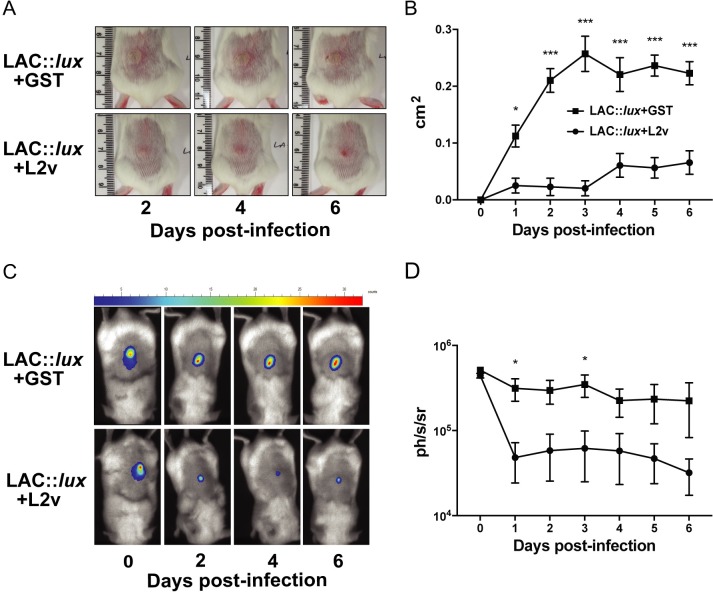
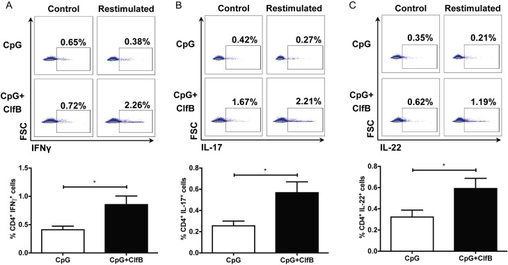
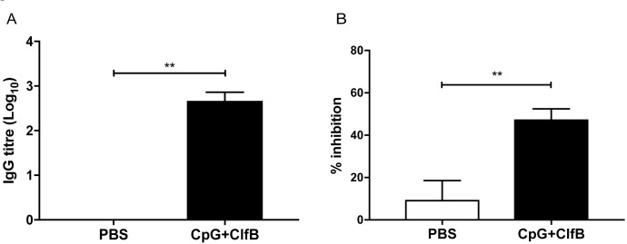
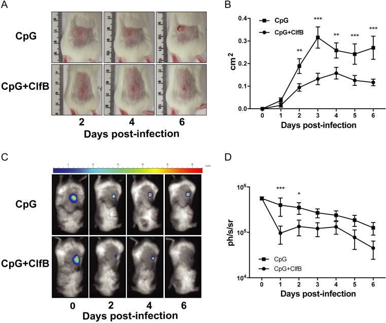
Staphylococcus aureus expresses a number of cell wall-anchored proteins that mediate adhesion and invasion of host cells and tissues and promote immune evasion, consequently contributing to the virulence of this organism. The cell wall-anchored protein clumping factor B (ClfB) has previously been shown to facilitate S. aureus nasal colonization through high affinity interactions with the cornified envelope in the anterior nares. However, the role of ClfB during skin and soft tissue infection (SSTI) has never been investigated. This study reveals a novel role for ClfB during SSTIs. ClfB is crucial in determining the abscess structure and bacterial burden early in infection and this is dependent upon a specific interaction with the ligand loricrin which is expressed within the abscess tissue. Targeting ClfB using a model vaccine that induced both protective humoral and cellular responses, leads to protection during S. aureus skin infection. This study therefore identifies ClfB as an important antigen for future SSTI vaccines.


Conflict of interest statement

The authors have declared that no competing interests exist.

Figures

Fig 1
Fig 1. ClfB contributes to virulence during S. aureus SSTI.
BALB/c mice were infected subcutaneously with 2x10⁷ CFU S. aureus LAC::lux or LAC::lux ΔclfB and abscess lesion size and bacterial burden was measured. Representative lesions from the dorsal area of mice from each group are shown (A) and results are expressed as total lesion size (cm2) ± SEM (B). Bioluminescence imaging was carried out using a Photon Imager and representative in vivo bioluminescence images are shown (C). Results are expressed as mean total photon flux (photons per second per steradian) ± SEM (D). Bacterial burden in the skin was assessed by viable counting on day 3 (E) and day 6 (F) post-infection. Results are expressed as Log10 CFU/mg. n = 9–10 per group. Data pooled from 2 independent experiments. Two-way ANOVA with Bonferroni post-test (B) and Mann-Whitney U test (E, F) used to analyse differences between groups. * P < 0.05, ** P < 0.01, *** P < 0.001.
Fig 2
Fig 2. ClfB contributes to abscess structure and size during S. aureus SSTI.
BALB/c mice were infected subcutaneously with 2x10⁷ CFU S. aureus LAC::lux or LAC::lux ΔclfB and abscess tissue was excised at 12, 48 and 96 hours. Tissue was fixed, embedded in paraffin wax and sectioned before haematoxylin and eosin staining was performed. Representative sections from each group are shown (A). White arrows indicate the lack of defined abscess wall structure. Tissue sections were scored (double blind) for histology score (B). Abscess area was computed using ImageJ software and the areas were scored accordingly (C). Results expressed as mean score ± SEM. n = 6 per group. Data pooled from 2 independent experiments. Two-way ANOVA with Bonferroni post-test used to analyze differences between groups (B, C). * P < 0.05, ** P < 0.01.
Fig 3
Fig 3. Loricrin in present within the skin abscess tissue of LAC::lux infected mice.
BALB/c mice were infected subcutaneously with 2x10⁷ CFU S. aureus LAC::lux and abscess tissue was excised at 48h post-infection. Tissue was fixed, embedded in paraffin wax and sectioned before anti-loricrin staining (dark brown) was carried out. Low power field of the entire abscess structure is shown (A). Relevant tissue sections are shown in high power fields: skin epidermal layer (B) and abscess wall (C). Black arrows indicate the presence of loricrin. Representative images of n = 3 stained sections.
Fig 4
Fig 4. Loricrin is an important ligand during S. aureus SSTI.
Wild-type FVB (WT) and Lor-/- mice were infected subcutaneously with 2x10⁷ CFU S. aureus LAC::lux or LAC::lux ΔclfB and abscess lesion size and bacterial burden was measured. Representative lesions from the dorsal area of mice from each group are shown (A) and results are expressed as total lesion size (cm2) ± SEM (B). Bioluminescence imaging was carried out using a Photon Imager and representative in vivo bioluminescence images are shown (C). Results are expressed as mean total photon flux (photons per second per steradian) ± SEM (D). Bacterial burden in the skin was assessed by viable counting on day 6 (E) post-infection. Results are expressed as Log10 CFU/mg. n = 5 per group. Tissue sections from WT and Lor-/- infected mice 12 h post-infection were stained with haematoxylin and eosin (F). Representative sections from each group are shown. Tissue sections were scored (double blind) for histology score (G). Abscess area was computed using ImageJ software and the areas were scored accordingly (H). Results expressed as mean score ± SEM. n = 4 per group. Two-way ANOVA with Bonferroni post-test (B, D), one-way ANOVA with Tukey post-test (E) and Mann-Whitney U test (G, H) used to analyze differences between groups. * P < 0.05, ** P < 0.01.
Fig 5
Fig 5. In vivo inhibition of ClfB-loricrin interaction leads to reduced abscess area and bacterial burden.
BALB/c mice were infected subcutaneously with 2x10⁷ CFU S. aureus LAC::lux pre-incubated with loricrin loop 2 region (L2v) or GST (24 μM) and abscess lesion size and bacterial burden was measured. Representative lesions from the dorsal area of mice from each group are shown (A) and results are expressed as total lesion size (cm2) ± SEM (B). Bioluminescence imaging was carried out using a Photon Imager and representative in vivo bioluminescence images are shown (C). Results are expressed as mean total photon flux (photons per second per steradian) ± SEM (D). n = 8 per group. Data pooled from 2 independent experiments. Two-way ANOVA with Bonferroni post-test used to analyze differences between groups. * P < 0.05, *** P < 0.001.
Fig 6
Fig 6. Vaccination with ClfB in combination with CpG leads to cellular immune responses.
BALB/c mice were vaccinated subcutaneously with CpG (50μg/mouse) alone or in combination with ClfB (5μg/mouse) on day 0, 14, 28. Antigen-specific cellular immune responses were measured on day 42 by ex vivo stimulation of inguinal lymph node cells with ClfB (10μg/ml). The percentage of CD4+IFN𝛾+ (A), CD4+IL-17+ (B) and CD4+IL-22+ (C) cells within the CD45+CD3+ population was assessed by flow cytometry. Results expressed as mean percentage ± SEM. n = 5–6 per group. Mann-Whitney U test used to analyze differences between groups. * P < 0.05.
Fig 7
Fig 7. Vaccination with ClfB in combination with CpG induces functional blocking ClfB-specific antibodies.
BALB/c mice were vaccinated subcutaneously with PBS or CpG in combination with ClfB (5μg/mouse) on day 0, 14, 28. Sera were collected on day 42 to assess antigen-specific humoral immune responses. ClfB-specific antibody titers were determined by ELISA and results are expressed as Log10 IgG titer (A). The presence of neutralizing antibodies was determined by measuring the ability of sera to inhibit the binding of L. lactis expressing ClfB (pKS80::clfB) to immobilised loricrin (B). n = 5 per group. Mann-Whitney U test used to analyze differences between groups. ** P < 0.01.
Fig 8
Fig 8. Vaccination with ClfB in combination with CpG offers protection against S. aureus SSTI.
BALB/c mice were vaccinated subcutaneously with CpG (50μg/mouse) alone or in combination with ClfB (5μg/mouse) on day 0, 14, 28. On day 42, mice were infected subcutaneously with 2x10⁷ CFU S. aureus LAC::lux and abscess lesion size and bacterial burden was measured. Representative lesions from the dorsal area of mice from each group are shown (A) and results are expressed as total lesion size (cm2) ± SEM (B). Bioluminescence imaging was carried out using a Photon Imager and representative in vivo bioluminescence images are shown (C). Results are expressed as mean total photon flux (photons per second per steradian) ± SEM (D). Results are expressed as Log10 CFU/mg. n = 10 per group. Data pooled from 2 independent experiments. Two-way ANOVA with Bonferroni post-test used to analyze differences between groups. * P < 0.05, ** P < 0.01, *** P < 0.001.

Similar articles

Cited by

References

    1. McCaig LF, McDonald LC, Mandal S, Jernigan DB. Staphylococcus aureus-associated skin and soft tissue infections in ambulatory care. Emerging infectious diseases. 2006;12(11):1715–23. Epub 2007/02/08. 10.3201/eid1211.060190 ; PubMed Central PMCID: PMCPmc3372331. - DOI - PMC - PubMed
    1. Singer AJ, Talan DA. Management of skin abscesses in the era of methicillin-resistant Staphylococcus aureus. The New England journal of medicine. 2014;370(11):1039–47. Epub 2014/03/14. 10.1056/NEJMra1212788 . - DOI - PubMed
    1. Hersh AL, Chambers HF, Maselli JH, Gonzales R. National trends in ambulatory visits and antibiotic prescribing for skin and soft-tissue infections. Archives of internal medicine. 2008;168(14):1585–91. Epub 2008/07/30. 10.1001/archinte.168.14.1585 . - DOI - PubMed
    1. Mansour SC, Pletzer D, de la Fuente-Nunez C, Kim P, Cheung GYC, Joo HS, et al. Bacterial Abscess Formation Is Controlled by the Stringent Stress Response and Can Be Targeted Therapeutically. EBioMedicine. 2016;12:219–26. Epub 2016/10/26. 10.1016/j.ebiom.2016.09.015 - DOI - PMC - PubMed
    1. David MZ, Daum RS. Community-associated methicillin-resistant Staphylococcus aureus: epidemiology and clinical consequences of an emerging epidemic. Clinical microbiology reviews. 2010;23(3):616–87. Epub 2010/07/09. 10.1128/CMR.00081-09 ; PubMed Central PMCID: PMCPmc2901661. - DOI - PMC - PubMed

Publication types

MeSH terms