. 2019 Apr 22;15(4):e1007717.
doi: 10.1371/journal.ppat.1007717. eCollection 2019 Apr.

Candida albicans induces mucosal bacterial dysbiosis that promotes invasive infection

Affiliations

Candida albicans induces mucosal bacterial dysbiosis that promotes invasive infection

Martinna Bertolini et al. PLoS Pathog. .

Abstract

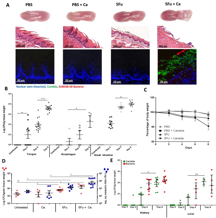
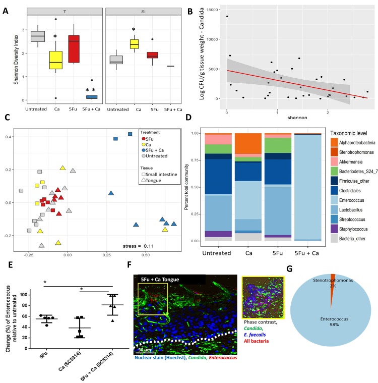
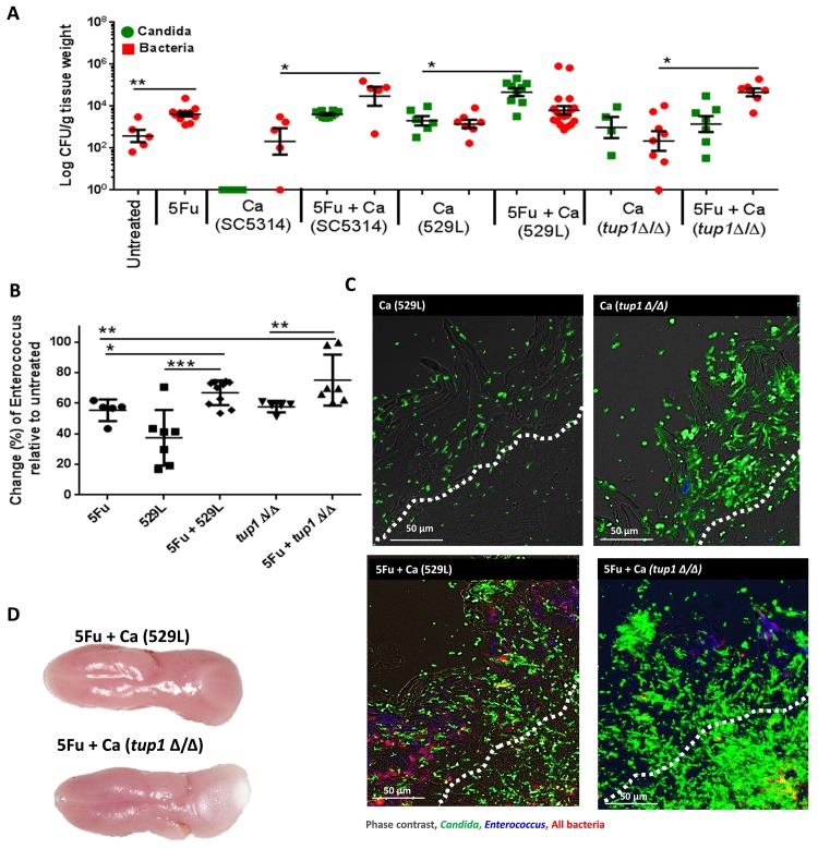
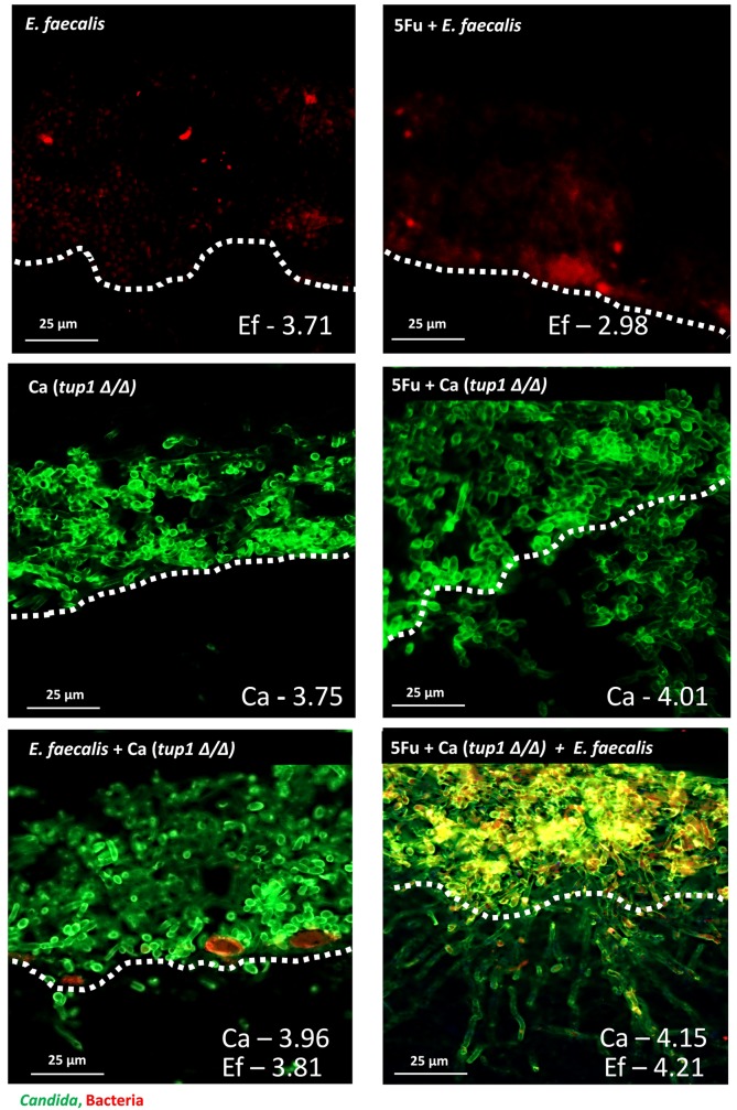
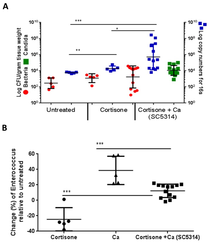
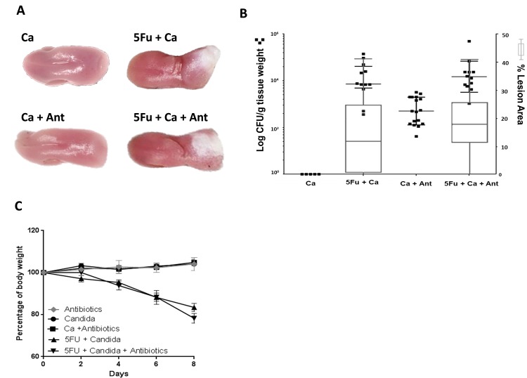
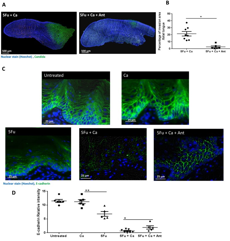
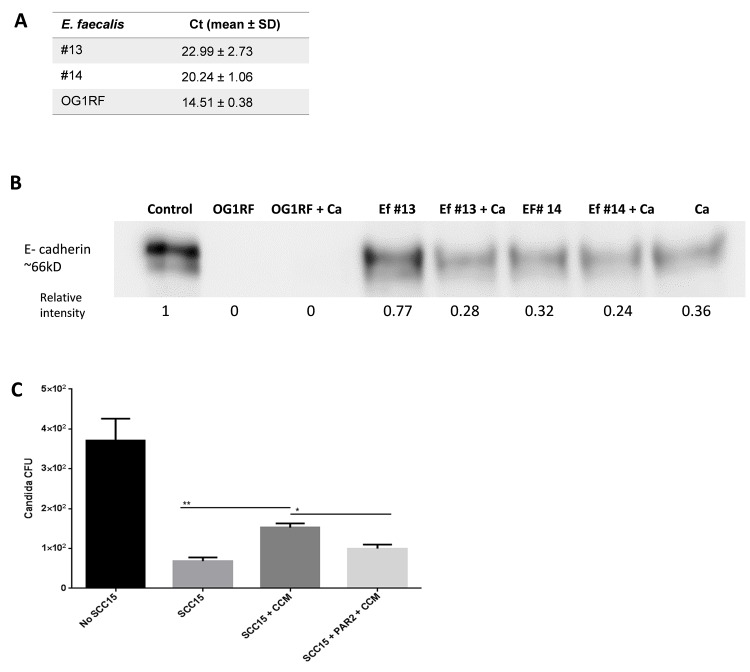
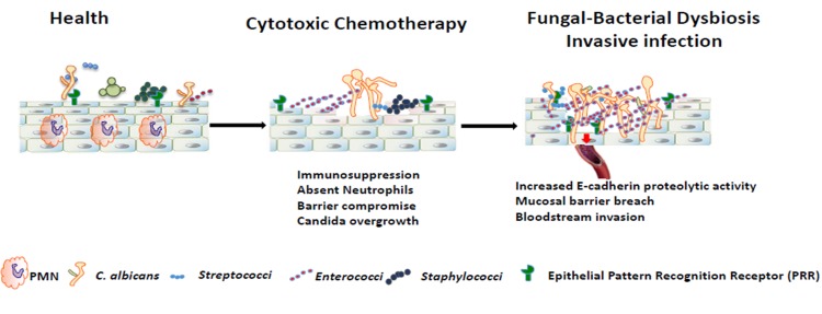
Infectious complications are a common cause of morbidity and mortality in cancer patients undergoing chemotherapy due to increased risk of oral and gastrointestinal candidiasis, candidemia and septicemia. Interactions between C. albicans and endogenous mucosal bacteria are important in understanding the mechanisms of invasive infection. We published a mouse intravenous chemotherapy model that recapitulates oral and intestinal mucositis, and myelosuppression in patients receiving 5-fluorouracil. We used this model to study the influence of C. albicans on the mucosal bacterial microbiome and compared global community changes in the oral and intestinal mucosa of the same mice. We validated 16S rRNA gene sequencing data by qPCR, in situ hybridization and culture approaches. Mice receiving both 5Fu and C. albicans had an endogenous bacterial overgrowth on the oral but not the small intestinal mucosa. C. albicans infection was associated with loss of mucosal bacterial diversity in both sites with indigenous Stenotrophomonas, Alphaproteobacteria and Enterococcus species dominating the small intestinal, and Enterococcus species dominating the oral mucosa. Both immunosuppression and Candida infection contributed to changes in the oral microbiota. Enterococci isolated from mice with oropharyngeal candidiasis were implicated in degrading the epithelial junction protein E-cadherin and increasing the permeability of the oral epithelial barrier in vitro. Importantly, depletion of these organisms with antibiotics in vivo attenuated oral mucosal E-cadherin degradation and C. albicans invasion without affecting fungal burdens, indicating that bacterial community changes represent overt dysbiosis. Our studies demonstrate a complex interaction between C. albicans, the resident mucosal bacterial microbiota and the host environment in pathogenesis. We shed significant new light on the role of C. albicans in shaping resident bacterial communities and driving mucosal dysbiosis.


Conflict of interest statement

N/A

Figures

Fig 1
Fig 1. Effect of 5Fu administration on Candida albicans and endogenous bacterial burdens.
A: Mucosal biofilm forming on tongues excised 8 days post-treatment. The group receiving 5Fu (50mg/kg, IV, every 48 hours) + C. albicans SC5314 (5Fu+Ca) had a thick mucosal biofilm covering the posterior tongue surface. Middle panel includes H&E-stains showing reduced epithelial thickness, papillary atrophy, desquamation and erosion in the 5Fu group and deep epithelial ulcerations in the 5Fu+Ca group. Lower panel shows immunofluorescence combined with fluorescence in situ hybridization (immuno-FISH) to simultaneously visualize C. albicans and bacterial commensals in tongue biofilms. C. albicans stained with a polyclonal anti-Candida antibody (green), commensal bacteria stained with EUB338-Alexa 546 probe (red) and cell nuclei counterstained with Hoechst 33258 (blue). B: Recovery of C. albicans from tongues, esophagus and small intestines in control untreated mice, and in mice receiving 5Fu every 48h with C. albicans SC5314 added daily in the drinking water. Shown are colony-forming unit (CFU) counts from organs harvested at baseline (uninfected), then 2, 6 and 8 days after the first 5Fu injection. Tissues were weighed, homogenized, serially diluted and plated for counts on Sabouraud Dextrose Agar containing chloramphenicol. Similar counts of C. albicans were obtained in CHROMagar Candida media with no other species identified (not shown). CFU assays showed that fungal burdens increased significantly over time in all tissues. Log CFU counts/gm of tissue are shown from 2 independent mouse experiments, with 5–15 mice per group; bars represent means ± SEM. *p<0.05, **p<0.0001. C: Body weight loss during the eight-day experimental period, expressed as percentage of initial weight (day 0) in 5–10 animals per group from 1–2 independent experiments. Error bars represent SEM. Mice receiving 5Fu every 48h lost weight slowly over time, while mice receiving 5Fu every 48 hours with C. albicans SC5314 added daily in the drinking water reached 20% body of total weight loss by day 8 (p<0.05 for a comparison with the 5Fu group for d8). D: Tongue bacterial loads compared among untreated mice, mice inoculated with C. albicans SC5314 (Ca), mice receiving 5Fu alone, and mice receiving both 5Fu and C. albicans SC5314 (5Fu+Ca) daily in the drinking water. Mice were sacrificed 8 days later. Tongues were weighed, homogenized, serially diluted and plated and results expressed as CFU counts/gm of tissue (left Y-axis, red dots). The total bacterial biomass (log 16S rRNA gene copy numbers/gm of tissue) was also quantified by real-time qPCR (right Y-axis, blue squares). Both assays showed a significant increase in endogenous tongue bacteria in 5Fu treated mice when compared to untreated and a further increase in the 5Fu + C. albicans group, which was significantly higher than the 5Fu group (p = 0.008). Data shown are from 1–2 independent mouse experiments, with 4–10 mice per group; bars represent means ± SEM. *p<0.01, p**<0.005, ***p<0.0005. E: Recovery of C. albicans and endogenous bacteria from kidneys and livers in mice receiving 5Fu and C. albicans SC5314 daily in the drinking water. Mice were sacrificed on days indicated. Kidneys and livers were weighed, homogenized, serially diluted and plated for CFU counts. Results show a time-dependent increase in fungal and bacterial dissemination to both organs. Log CFU counts/gm of tissue are shown from 2 independent mouse experiments, with 5–10 mice per group; bars represent means ± SEM. *p<0.0001, **p<0.0005.
Fig 2
Fig 2. Mucosa-associated bacterial microbiota profiling.
A: Microbial DNA was extracted from tongues (T) and small intestines (SI) of the same mice on day 8. The V4 hypervariable region of the 16S rDNA gene was amplified and sequenced. Box plot showing Shannon Diversity Index in untreated mice, mice receiving C. albicans SC5314 daily in the drinking water (Ca), mice receiving 5Fu alone and a combination of the two (5Fu+Ca). Mean diversity index values are shown from 5 mice in each group. In mice inoculated with C. albicans alone (Ca) bacterial diversity decreased in the tongue but increased in the small intestine mucosa (*p<0.05 for a comparison to untreated groups). For mice receiving 5Fu and C. albicans (5Fu+Ca) there was a significant further reduction in bacterial diversity in tongue but not small intestinal tissues (**p<0.001). B: Linear regression analysis of C. albicans CFUs plotted against the Shannon Diversity Index of the bacterial communities in the same tongues. Data include all mice receiving C. albicans SC5314 with or without chemotherapy, at all time points (5 mice/group, t = 2, 6 or 8 days, n = 30). This analysis showed higher C. albicans burdens correlating with lower bacterial diversity (R2 = 0.17, p<0.05). C: Beta diversity assessed by nonmetric multidimensional scaling (NMS) based on Bray-Curtis dissimilarities among four treatment groups. Shown are community structures in the untreated control, C. albicans alone, and the two chemotherapy groups (Untreated: grey, Ca: yellow 5Fu: red, 5Fu+Ca: blue; n = 5 mice/group) in tongues (triangles) and small intestines (squares) of the same mice. Results represent community structure differences at the end of the experimental period (day 8). Microbial communities clustered by type of treatment, indicating a significant effect of C. albicans in 5Fu treated mice. Samples in this group also clustered by site, indicating that the two sites harbor microbiomes with distinct global community structures and composition by day 8. The type of treatment explained 16% of the variability (p<0.01), whereas the site explained 8.5% of the variability (p<0.01) among samples. D: Mean relative abundance of OTU sequences assigned to one of the top 10% prominent taxa identified on mouse tongues, in each of the four treatment groups, at the end of the experimental period (day 8, n = 5 mice/group). Enterococcus was the most abundant taxon in both C. albicans-inoculated groups. However, relative abundance of this genus was more extensive in mice that also received chemotherapy, in which they represented ~98% of the total bacterial community. E: Validation of the bacterial microbiome sequencing data by qPCR. Genus level quantification was performed for Enterococcus on tongue tissues by qPCR. Data represent change in percentage of Enterococcus load in 5Fu, Ca (C. albicans SC5314) and 5Fu + Ca (SC5314) groups compared to untreated control mice. Mice receiving both 5Fu and C. albicans had significantly higher Enterococcus increase when compared to all other groups. Results are shown from 5 mice per group; bars represent means ± SEM. *p<0.001. F: Tongue tissue section from mice receiving 5Fu and C. albicans SC5314 for 8 days. Tissue sections were stained by immuno-FISH to simultaneously visualize C. albicans and endogenous bacteria. Left panel shows staining with the Enterococcus/Lactobacillus specific probe LAB158 (red), anti-Candida-FITC polyclonal antibody (green) and cell nuclei counterstained with Hoechst 33258 (blue). Note Candida invasion in the submucosal compartment (bellow the white dotted line). On the right panel immuno-FISH staining of a serial section from the same tissue is shown representing the biofilm area in yellow. Section was triple-stained with the all bacteria probe EUB338 (red), the Enterococcus faecalis specific probe ENFL84 (blue), and anti-Candida antibody (green). Note the almost complete overlap of the blue and red signals resulting in blue-purple staining bacteria representing E. faecalis. G: Identification of bacteria isolated from the tongues of mice after 8 days of receiving 5Fu and C. albicans SC5314. The V4 variable region of the 16S rDNA gene was amplified and sequenced by Illumina. 98% of the OTU sequences from five isolates aligned with the genus Enterococcus. All isolates were identified as E. faecalis with species-specific primers and PCR.
Fig 3
Fig 3. Effects of different C. albicans strains and 5Fu on oral bacteria.
A: Recovery of C. albicans (green) and total cultivable bacteria (red) from tongues of untreated mice, mice receiving 5Fu, mice receiving C. albicans (Ca) and a combination (5Fu+Ca). C. albicans strains SC5314, 529L or the tup1Δ/Δ deletion mutant were used in these experiments. Tongues were weighed, homogenized, serially diluted and plated for CFU counts. Strains 529L and tup1Δ/Δ were able to stably colonize the tongue mucosa in the absence of 5Fu, however, this was not associated with increased oral bacterial loads. On the other hand the combination of 5Fu with both strains led to increased bacterial loads. Log CFU counts/gm of tissue are shown from 2 independent mouse experiments, with 4–8 mice per group; bars represent means ± SEM. *p<0.01, **p<0.0001. B: Tongue-associated Enterococcus biomass as assessed by genus-level qPCR. Data represent percentage change of Enterococcus genome copy numbers relative to untreated mice. Groups included untreated mice, mice receiving 5Fu, strain 529L alone, strain 529L + 5Fu, the tup1Δ/Δ deletion mutant alone and the tup1Δ/Δ deletion mutant + 5Fu. All treatments led to an increase in the enterococcal biomass, however mice receiving both 5Fu and C. albicans had the highest increase. Results shown are from 5–8 mice per group in two experiments; bars represent means ± SEM. *p<0.05, **p<0.005, ***p<0.002. C: Tongue tissue sections from mice receiving C. albicans 529L or tup1Δ/Δ deletion mutant in the absence (left panel) or presence (right panel) of concomitant 5Fu chemotherapy for 8 days. Tissue sections were triple-stained with the all bacteria probe EUB338 (red), the E. faecalis specific probe ENFL84 (blue), and anti-Candida antibody (green). Enterococcal signal shown as purple-blue is clearly visible in mice receiving 5Fu with both strains. In these mice there was fungal invasion into the oral submucosa (bellow the white dotted line), which was more pronounced with the tup1Δ/Δ mutant. D: Representative tongues excised from mice receiving 5Fu with strain 529L (top) or the tup1Δ/Δ deletion mutant (bottom), 8 days after infection. Note the white biofilm area in the posterior surface with the tup1Δ/Δ deletion mutant.
Fig 4
Fig 4. Role of mucosal injury in biofilm growth and invasion using an organotypic model.
An E. faecalis isolate was inoculated alone or in combination with C. albicans strain tup1Δ/Δ. Biofilms are shown growing on mucosal constructs that had been pretreated with 10 μM 5Fu for 16 h (right panel) or on untreated controls (left panel). Tissues were split in half with one half processed for CFU counts and one half for histologic processing and staining. For CFU counts superficially growing biofilms were rinsed off and tissues were weighed, homogenized, and plated. Log10 CFU counts corresponding to each tissue and each organism are shown on the lower right. Unwashed tissues were stained with immuno-FISH to simultaneously visualize C. albicans and Enterococcus. C. albicans was stained with a polyclonal anti-Candida antibody (green), and E. faecalis was stained with EUB338-Alexa 546 probe (red), and areas of co-localization show in yellow. Fungal invasion was more pronounced in 5Fu-treated tissues infected with both organisms and corresponded to bright yellow areas of bacterial and fungal co-localization right above the mucosal breach point. Dotted lines demarcate the start of the submucosal compartment. One of three representative tissues is shown/condition.
Fig 5
Fig 5. Effect of cortisone immunosuppression and C. albicans infection on oral bacteria.
Mice were immunosuppressed by subcutaneous injection with cortisone acetate and inoculated with C. albicans SC5314 as described in methods. A: Tongue mucosa-associated bacterial loads in untreated, cortisone-treated, and cortisone-treated C. albicans-infected mice. Tongues were weighed, homogenized, serially diluted and plated for Candida and bacterial burdens. Fungal burdens in infected mice are expressed as log CFU/gm of tissue (green squares, left Y axis). Results for bacteria are expressed as CFU/gm of tissue (left Y-axis, red dots) and log 16S rRNA gene copy numbers/gm of tissue (right Y-axis, blue squares). Bacterial biomass increased with cortisone treatment, but a further significant increase was noted in the cortisone treated + C. albicans group. Data shown are from 2 independent mouse experiments, with 5–10 mice per group; bars represent means ± SEM. *p<0.05, **p<0.005, ***p<0.002. B: Genus level quantification of Enterococcus on tongue tissues by qPCR. Data represent change in percentage of Enterococcus load in all groups over untreated control mice. Mice receiving cortisone had a reduction in Enterococcus burdens while C. albicans in cortisone-treated mice raised the burdens close to untreated controls. Results are shown from 5–13 mice per group; bars represent means ± SEM. *p<0.001.
Fig 6
Fig 6. Effect of antibiotic treatment on oral fungal burdens and biofilm surface area in 5Fu-treated mice.
Antibiotics were started three days prior to 5Fu-treatment and continued for 8 days. A: Excised tongues after 8 days of C. albicans SC5314 inoculation. Groups receiving 5Fu with C. albicans SC5314 (5Fu+Ca) had thick mucosal biofilms covering the posterior tongue surface regardless of antibiotics (Ant) treatment. No biofilms formed in C. albicans-inoculated healthy mice with (Ca+Ant) or without (Ca) antibiotic treatment. B: Relationship between C. albicans burdens and biofilm surface area. Figure depicts log fungal CFUs/gm of tissue (dot plot, left Y axis) and corresponding biofilm lesion area (box and whiskers plot, right Y axis) in tongues of four groups of mice (day 8). Mice received C. albicans SC5314 in the drinking water alone (Ca), 5Fu with C. albicans (5Fu+Ca), C. albicans with antibiotics (Ca+Ant) or combination of the three (5Fu+Ca+Ant). Tissues were homogenized, serially diluted and plated for CFU counts. Biofilms were digitally photographed, images were analyzed by Image J and results were expressed as the percentage of dorsal tongue area covered by visible biofilm (white area). In groups treated with 5Fu there were no differences in fungal burdens (p = 0.53) or biofilm area (p = 0.25) between mice receiving antibiotics or not. In mice not receiving 5Fu, antibiotic treatment led to increased C. albicans burdens (p<0.005) but there was no biofilm lesion on the tongue surface indicating that in healthy mice increased fungal burdens are not sufficient for virulence. Data are from 2 independent mouse experiments, with 5–8 mice per group; bars represent means ± SEM. C: Body weight loss in each group during the eight-day infection period, expressed as percentage of initial weight (day 0) in 5–10 animals per group from 2 independent experiments. Error bars represent SEM. Candida-Infected mice receiving 5Fu with or without antibiotics lost 20% of total body weight by day 8.
Fig 7
Fig 7. Enterococcus depletion by antibiotic treatment significantly reduced oral mucosal C. albicans invasion and E-cadherin degradation from epithelial adherens junctions.
A: Full tongue sagittal section scans from mice receiving 5Fu with C. albicans SC5314 (5Fu+Ca) or mice additionally receiving antibiotics (5Fu+Ca+Ant) 8 days after initiation of 5Fu and fungal inoculation. Tongues were stained by immunofluorescence with an anti-Candida-FITC polyclonal antibody (green), and counterstained with Hoechst 33258 (blue) to visualize cell nuclei. Note the reduction in C. albicans invasion into the submucosal tissues with antibiotic treatment. B: Tongues stained as above were analyzed by Image J and the percentage of area invaded by fungi (green over blue signal) was calculated. Data is from 2 independent mouse experiments, with 3–5 mice per group; bars represent means ± SD. *p<0.001. C: Tongue tissue sections from untreated mice, mice receiving 5Fu alone, mice inoculated with C. albicans SC5314 alone (Ca), mice receiving 5Fu and C. albicans (5Fu+Ca) or mice additionally receiving antibiotics (5Fu+Ca+Ant) sacrificed 8 days after initiation of 5Fu and fungal inoculation. Tongues were stained by immuno-fluorescence for E-cadherin (green) and counterstained with nucleic acid stain Hoechst 33258 (blue). E-cadherin signal was intact in untreated mice and healthy mice inoculated with C. albicans, indicating integrity of adherens junctions. Signal was reduced in all other groups, with the greatest reduction in mice receiving 5Fu and C. albicans. D: E-cadherin staining was analyzed by ImageJ and mean intensity of the green signal per mm2 was calculated in tissue sections from 6 mice/group. Bars represent means ± SD. *p<0.05, **p<0.005.
Fig 8
Fig 8. Enterococcus isolates from oral dysbiotic communities are able to degrade E-cadherin and promote epithelial permeability.
A: Analysis of E. faecalis gelE gene expression by quantitative RT-PCR. Comparison of gene expression levels in stationary phase overnight cultures of isolates #13 and #14, with strain OG1RF used as a positive control. Relative RNA levels were calculated using the DDCt method and recA gene expression was used as an internal control to normalize RNA concentration. Data were expressed as mean threshold cycle (Ct) ± SD of duplicate samples in at least three independent cultures. B: E-cadherin degradation assay. Concentrated conditioned media from stationary phase cultures of E. faecalis isolates #13 and #14 were incubated with 3 μg of recombinant E-cadherin for 1 hour, with or without conditioned media from C. albicans SC5314. E. faecalis strain OG1RF was used as a positive control. Western blot shows reduction of E-cadherin signal with both isolated strains and complete degradation of the protein with strain OG1RF. Numbers under each lane represent relative image density as measured with the Image J software. One representative of three experiments is shown. C: Migration of C. albicans (SC5314) across confluent SCC15 cell monolayers in a transwell assay. Fungi translocating to the lower chamber after 4 hours incubation were quantified by plating the media for CFUs. C. albicans migration was significantly greater when monolayers were apically pretreated with E. faecalis Concentrated Cell Conditioned Media (CCM) compared to untreated or CCM+PAR2 inhibitor pretreated controls. Results are from 2 independent experiments with 3 technical replicates in each. Data are expressed as mean values and SEM. *p<0.05, **p<0.0001.
Fig 9
Fig 9. Model of oral mucosal candidiasis in chemotherapy.
Cytotoxic chemotherapy promotes a dysbiotic state characterized by overgrowth of C. albicans and certain resident bacterial species. Bacterial proteolytic activity promotes mucosal barrier breach by C. albicans.

Similar articles

Cited by

References

    1. Lalla RV, Latortue MC, Hong CH, Ariyawardana A, D'Amato-Palumbo S, Fischer DJ, Martof A, Nicolatou-Galitis O, Patton LL, Elting LS, Spijkervet FK, Brennan MT; Fungal Infections Section, Oral Care Study Group, Multinational Association of Supportive Care in Cancer (MASCC)/International Society of Oral Oncology (ISOO). A systematic review of oral fungal infections in patients receiving cancer therapy. Support Care Cancer. 2010. August;18(8):985–92. 10.1007/s00520-010-0892-z - DOI - PMC - PubMed
    1. Peterson DE, Boers-Doets CB, Bensadoun RJ, Herrstedt J; ESMO Guidelines Committee. Management of oral and gastrointestinal mucosal injury: ESMO Clinical Practice Guidelines for diagnosis, treatment, and follow-up. Ann Oncol. 2015. September;26 Suppl 5:v139–51. 10.1093/annonc/mdv202 Epub 2015 Jul 4. - DOI - PubMed
    1. Cole GT, Halawa AA, Anaissie EJ. The role of the gastrointestinal tract in hematogenous candidiasis: from the laboratory to the bedside. Clinical infectious diseases: an official publication of the Infectious Diseases Society of America. 1996;22 Suppl 2:S73–88. - PubMed
    1. Jarvis WR. Epidemiology of nosocomial fungal infections, with emphasis on Candida species. Clinical infectious diseases: an official publication of the Infectious Diseases Society of America. 1995;20(6):1526–30. - PubMed
    1. Teoh F, Pavelka N. How Chemotherapy Increases the Risk of Systemic Candidiasis in Cancer Patients: Current Paradigm and Future Directions. Pathogens. 2016. January 15;5(1). pii: E6 10.3390/pathogens5010006 - DOI - PMC - PubMed

Publication types

MeSH terms