

Development of lacrimal gland inflammation in the mouse model of herpes stromal keratitis
- PMID: 31009613
- PMCID: PMC6570564
- DOI: 10.1016/j.exer.2019.04.022
Development of lacrimal gland inflammation in the mouse model of herpes stromal keratitis
Abstract
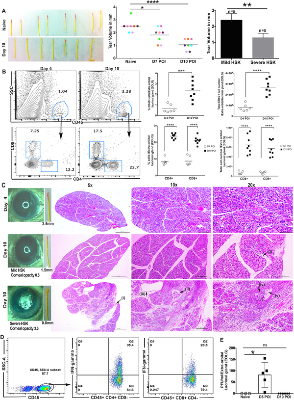
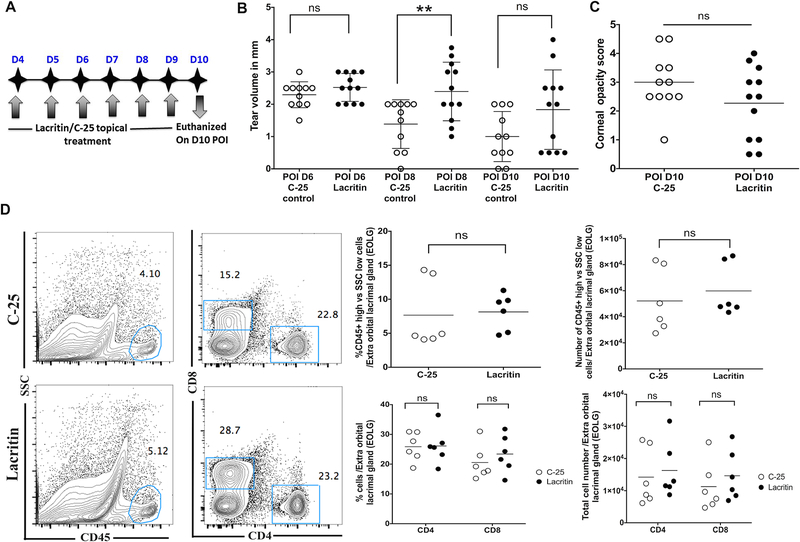
Herpes stromal keratitis (HSK) is a chronic immunoinflammatory condition which develops in response to recurrent herpes simplex virus-1 (HSV-1) infection of the cornea. Patients with HSK often demonstrate the concurrence of corneal desiccation and the loss of blink reflex. However, the relationship between severity of HSK, level of basal tears and inflammation of the lacrimal gland is mostly unexplored. In this study, we compared these variables in extraorbital lacrimal gland (EoLG) after corneal HSV-1 infection in the C57BL/6J mouse model. Our results showed a significant reduction in the volume of tears in infected eyes during the development of HSK. Extensive architectural damage to EoLG, presumably caused by a massive influx of interferon-gamma secreting T cells, was observed during clinical disease period of HSK. A positive correlation between the decrease in tear volume, severity of HSK and the damage to EoLG were evident in infected mice. The presence of infectious virus measured in EoLG during pre-clinical, but not clinical disease period of HSK, suggested that viral cytopathic effects are not the major contributors of extensive damage seen in EoLG. Furthermore, topical administration of lacritin peptide delayed but did not prevent the decrease in tears in HSV-1 infected mice, and had no significant effect in either reducing the severity of HSK or T cell infiltration in EoLG of infected mice. Together, our results showed an interplay between the severity of HSK, inflammation of EoLG, and the reduced level of tears after corneal HSV-1 infection.
Keywords: HSV-1; Lacrimal gland; Lacritin and T cells; Tears.
Copyright © 2019 Elsevier Ltd. All rights reserved.
Conflict of interest statement
Conflict of interest
GWL is cofounder of TearSolutions, Inc, that is currently testing the efficacy of a lacritin synthetic peptide for Sjogren’s syndrome dry eye in a phase 2 clinical trial. RLM is contractor of TearSolutions.
Figures
Similar articles
-
Herpes stromal keratitis erodes the establishment of tissue-resident memory T cell pool in HSV-1 infected corneas.Mucosal Immunol. 2025 Feb;18(1):188-204. doi: 10.1016/j.mucimm.2024.11.003. Epub 2024 Nov 22. Mucosal Immunol. 2025. PMID: 39581232 Free PMC article.
-
Sensory Nerve Retraction and Sympathetic Nerve Innervation Contribute to Immunopathology of Murine Recurrent Herpes Stromal Keratitis.Invest Ophthalmol Vis Sci. 2022 Feb 1;63(2):4. doi: 10.1167/iovs.63.2.4. Invest Ophthalmol Vis Sci. 2022. PMID: 35103749 Free PMC article.
-
Reversible nerve damage and corneal pathology in murine herpes simplex stromal keratitis.J Virol. 2014 Jul;88(14):7870-80. doi: 10.1128/JVI.01146-14. Epub 2014 Apr 30. J Virol. 2014. PMID: 24789786 Free PMC article.
-
[Research progress of corneal neovascularization in herpes stromal keratitis].Zhonghua Yan Ke Za Zhi. 2019 Dec 11;55(12):956-960. doi: 10.3760/cma.j.issn.0412-4081.2019.12.017. Zhonghua Yan Ke Za Zhi. 2019. PMID: 31874509 Review. Chinese.
-
Corneal lymphangiogenesis in herpetic stromal keratitis.Surv Ophthalmol. 2015 Jan-Feb;60(1):60-71. doi: 10.1016/j.survophthal.2014.06.001. Epub 2014 Jun 10. Surv Ophthalmol. 2015. PMID: 25444520 Free PMC article. Review.
Cited by
-
Lack of neonatal Fc receptor does not diminish the efficacy of the HSV-1 0ΔNLS vaccine against ocular HSV-1 challenge.Vaccine. 2021 Apr 28;39(18):2526-2536. doi: 10.1016/j.vaccine.2021.03.075. Epub 2021 Apr 1. Vaccine. 2021. PMID: 33814229 Free PMC article.
-
Dry eye disease caused by viral infection: Past, present and future.Virulence. 2024 Dec;15(1):2289779. doi: 10.1080/21505594.2023.2289779. Epub 2023 Dec 4. Virulence. 2024. PMID: 38047740 Free PMC article. Review.
-
CGRP Released by Corneal Sensory Nerve Maintains Tear Secretion of the Lacrimal Gland.Invest Ophthalmol Vis Sci. 2024 Apr 1;65(4):30. doi: 10.1167/iovs.65.4.30. Invest Ophthalmol Vis Sci. 2024. PMID: 38635244 Free PMC article.
-
Ocular Glands Become Infected Secondarily to Infectious Keratitis and Play a Role in Corneal Resistance to Infection.J Virol. 2019 Jul 30;93(16):e00314-19. doi: 10.1128/JVI.00314-19. Print 2019 Aug 15. J Virol. 2019. PMID: 31167909 Free PMC article.
-
Enterovirus A71 infection-induced dry eye-like symptoms by damaging the lacrimal glands.Front Cell Infect Microbiol. 2024 Apr 2;14:1340075. doi: 10.3389/fcimb.2024.1340075. eCollection 2024. Front Cell Infect Microbiol. 2024. PMID: 38628549 Free PMC article.
References
-
- Barron BA, Gee L, Hauck WW, Kurinij N, Dawson CR, Jones DB, Wilhelmus KR, Kaufman HE, Sugar J, Hyndiuk RA, et al., 1994. Herpetic Eye Disease Study. A controlled trial of oral acyclovir for herpes simplex stromal keratitis. Ophthalmology 101, 1871–1882. - PubMed
-
- Dallacasagrande V, Hirata H, Mizerska KK, van Kuppevelt TH, McKown RL, Laurie GW, 2016. Lacritin acutely enhances corneal nerve sensitivity to ocular surface dryness as a key stimulus for basal tear production: Implications for dry eye disease. Invest Ophth Vis Sci 57.
Publication types
MeSH terms
Substances
Grants and funding
LinkOut - more resources
Full Text Sources
