. 2019 Nov;44(12):2091-2098.
doi: 10.1038/s41386-019-0397-5. Epub 2019 Apr 22.

Molecular imaging identifies age-related attenuation of acetylcholine in retrosplenial cortex in response to acetylcholinesterase inhibition

Affiliations

Molecular imaging identifies age-related attenuation of acetylcholine in retrosplenial cortex in response to acetylcholinesterase inhibition

Theodosia Vallianatou et al. Neuropsychopharmacology. 2019 Nov.

Abstract

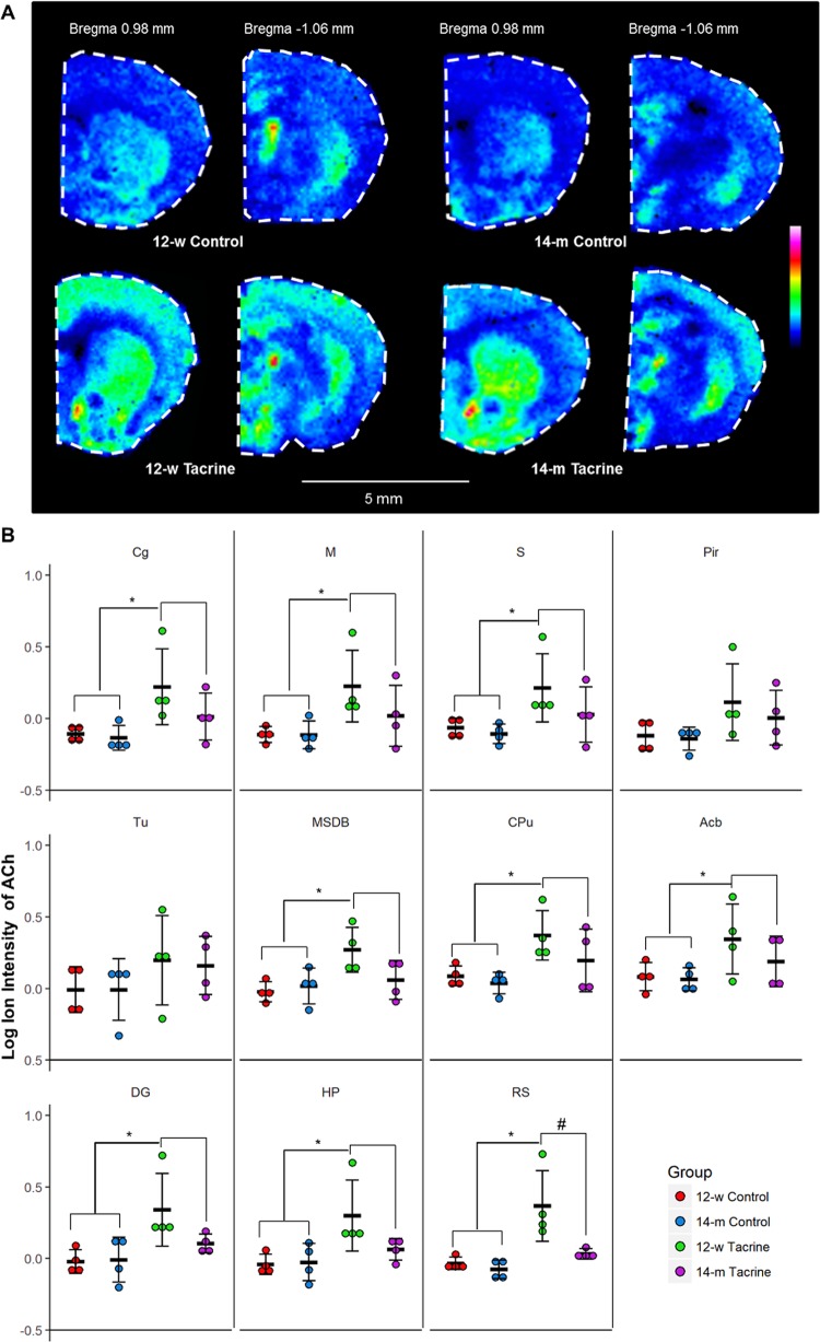
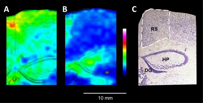
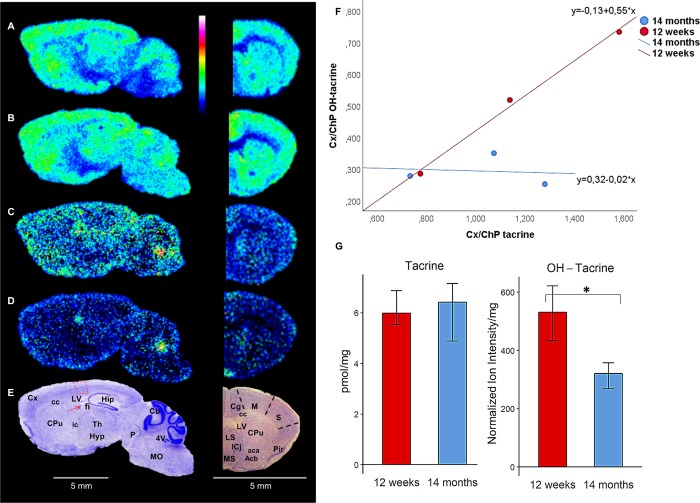
The neurotransmitter of the cholinergic system, acetylcholine plays a major role in the brain's cognitive function and is involved in neurodegenerative disorders. Here, we present age-related alterations of acetylcholine levels after administration of the acetylcholinesterase inhibitor drug tacrine in normal mice. Using a quantitative, robust and molecular-specific mass spectrometry imaging method we found that tacrine administration significantly raised acetylcholine levels in most areas of sectioned mice brains, inter alia the striatum, hippocampus and cortical areas. However, acetylcholine levels in retrosplenial cortex were significantly lower in 14-month-old than in 12-week-old animals following its administration, indicating that normal aging affects the cholinergic system's responsivity. This small brain region is interconnected with an array of brain networks and is involved in numerous cognitive tasks. Simultaneous visualization of distributions of tacrine and its hydroxylated metabolites in the brain revealed a significant decrease in levels of the metabolites in the 14-month-old mice. The results highlight strengths of the imaging technique to simultaneously investigate multiple molecular species and the drug-target effects in specific regions of the brain. The proposed approach has high potential in studies of neuropathological conditions and responses to neuroactive treatments.


Figures

Fig. 1
Fig. 1
Brain distribution of ACh in a tacrine-administered 14-m mouse sagittal brain tissue section. a MALDI-MSI of ACh (m/z 146.1176, scaled to 60% of max intensity and normalized to the internal standard) in a representative sagittal mouse brain section (lateral 1.7 mm) of a 14-m tacrine-administered animal at a lateral resolution of 60 μm. b The tissue section was subsequently washed and subjected to Nissl staining; brain structures of interest are annotated. ACh was highly localized in the Cx and Hip, which are areas receiving cholinergic innervation from the basal forebrain cholinergic nuclei. High levels of ACh were also detected in the striatum, i.e., CPu and Acb, in which cholinergic interneurons are present, as well as in the Tu. ACh abundance was also high in the AN and Th, receiving cholinergic input from the basal forebrain and upper brainstem, respectively, and the basal forebrain nuclei (DB), which are characterized by projecting cholinergic neurons, and in certain areas of the hindbrain and cerebellum, projected mainly by the mesencephalic cholinergic neurons. Abbreviations for gray matter areas: 5N motor trigeminal nucleus, 7N facial nucleus, Acb nucleus accumbens, AI agranular insular cortex, AN amygdalar nuclei, Cb cerebellum, CPu caudate putamen, Cx cerebral cortex, DB nucleus of diagonal band, GP globus pallidus, GrDG granular cell layer of dentate gyrus, Hip hippocampus, Hyp hypothalamus, LOT nucleus of the lateral olfactory tract, M motor cortex, Mitg microcellular tegmental nucleus, Pir piriform cortex, Post postsubiculum, PtA parietal association cortex, S somatosensory cortex, Sn substantia nigra, Sp5 spinal trigeminal nucleus, Th thalamus, Tu olfactory tubercle, V visual cortex, VP ventral pallidum. Abbreviations for white matter areas: 5n trigeminal (cranial) nerve, acp anterior commissure post limb, cc corpus callosum, fi fibria of hippocampus, ic internal capsule, lo lateral olfactory tract, mfb. medial forebrain bundle, opt optic tract. Abbreviations for ventricular areas: LV lateral ventricles 4V fourth ventricle
Fig. 2
Fig. 2
Molecule-specific imaging of ACh in coronal brain tissue sections. a MALDI-MSI of ACh in representative coronal brain sections of two brain levels (bregma 0.98 and −1.06 mm) [12] from 12-w to 14-m controls and tacrine-administered animals (images scaled to 40% of max intensity). b Dot plot of the log ion intensities of ACh in the investigated brain structures (n = 4). Error bars show 95% confidence interval. Significant difference (α = 0.05) between the two treatment groups is illustrated with (*). Significant difference (α = 0.05) between the 12-w tacrine and the 14-m tacrine groups in the area of the retrosplenial cortex is illustrated with (#). Abbreviations: Cg cingulate cortex, M motor cortex, S sensor cortex, Pir piriform, Tu olfactory tubercle, MSD medial septum/diagonal band, CPu caudate putamen, Acb nucleus accumbens, DG dentate gyrus, HP hippocampal proper, RS retrosplenial cortex
Fig. 3
Fig. 3
Age-related decrease of ACh concentration in retrosplenial cortex in response to acetylcholinesterase inhibition. a, b MALDI-MSI of ACh distribution in the retrosplenial cortex of 12-w and 14-m tacrine-dosed animals (100 μm lateral resolution, images scaled to 40% of max intensity). The retrosplenial cortex is highlighted by white dashed lines. The MALDI-MS and Nissl stained images are overlaid using the image fusion function implemented in msIQuant software [13] for better delineation of the brain structures. c The tissue section was washed and subjected to Nissl staining; brain structures of interest are annotated. Abbreviations: DG dentate gyrus, HP hippocampal proper, RS retrosplenial cortex
Fig. 4
Fig. 4
Brain distribution of tacrine and its hydroxylated metabolites after intraperitoneal administration (10 mg/kg). a, b Sagittal and coronal brain tissue sections imaged by MALDI-MS showing tacrine (m/z 199.123) in 12-w and 14-m mice, respectively (35% of max intensity). c, d Sagittal and coronal brain tissue sections imaged by MALDI-MS showing OH-tacrine (m/z 215.118) in 12-w and 14-m mice, respectively (20% of max intensity). Both analytes were imaged by MALDI-MS at a lateral resolution of 80 μm. e The sagittal and coronal brain tissue sections were subsequently washed and subjected to Nissl staining; brain structures of interest are annotated. Areas used for measuring the Cx/ChP ratio (ratio of a compound’s concentration in the somatosensory cortex to its concentration in the choroid plexus) in the sagittal brain tissue sections are highlighted in red. f Scatter plots of Cx/ChP ratios for tacrine (X-axis) and OH-tacrine (Y-axis) (n = 3). g Bar plot of the whole-brain concentration of tacrine (pmol/mg tissue) and normalized ion intensity of OH-tacrine (n = 4). Statistically significant differences (P < 0.05) are indicated with an asterisk (*). Abbreviations: Acb nucleus accumbens, Cb cerebellum, Cg cingulate cortex, ChP choroid plexus, CPu caudate putamen, Cx cortex, Hip hippocampus, Hyp hypothalamus, LS lateral septal nucleus, M motor cortex, MO medulla oblongata, MS medial septal nucleus, P pons, Pir piriform cortex, S somatocensory cortex, Th thalamus, aca anterior commissure anterior part, cc corpus callosum, f fimbria of the hippocampus, ic internal capsule, 4V fourth ventricle, LV lateral ventricle

Similar articles

Cited by

References

    1. Blokland A. Acetylcholine: a neurotransmitter for learning and memory? Brain Res Rev. 1995;21:285–300. - PubMed
    1. Euston DR, Gruber AJ, McNaughton BL. The role of medial prefrontal cortex in memory and decision making. Neuron. 2012;76:1057–70. - PMC - PubMed
    1. Gielow MR, Zaborszky L. The input-output relationship of the cholinergic basal forebrain. Cell Rep. 2017;18:1817–30. - PMC - PubMed
    1. Decker MW. The effects of aging on hippocampal and cortical projections of the forebrain cholinergic system. Brain Res Rev. 1987;12:423–38. - PubMed
    1. Bartus RT, Dean RL, III, Beer B, Lippa AS. The cholinergic hypothesis of geriatric memory dysfunction. Science. 1982;217:408–14. - PubMed

Publication types