

Herpes Simplex Virus Proctitis Masquerading as Rectal Cancer
- PMID: 31010103
- PMCID: PMC6630232
- DOI: 10.3390/diseases7020036
Herpes Simplex Virus Proctitis Masquerading as Rectal Cancer
Abstract
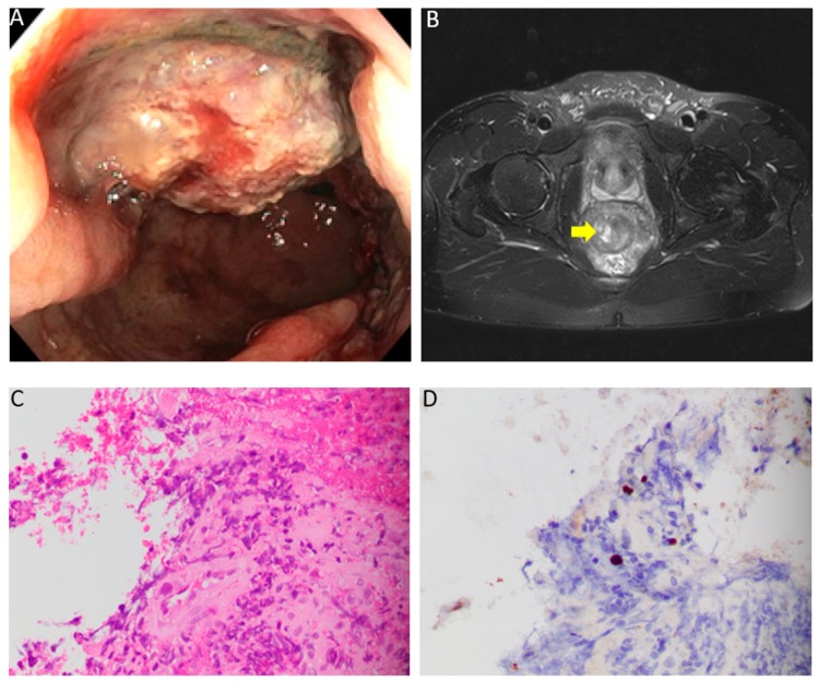
Herpes simplex virus (HSV) is the leading cause of proctitis in HIV-infected individuals. However, no cases of rectal masses secondary to HSV infection have been reported to date. Herein, we present the case of a 45-year-old man with HIV infection who developed rectal pain and bleeding, along with dysuria and voiding difficulty. Colonoscopy revealed proctitis and a rectal mass with features concerning for rectal cancer. Histologic sections of the rectal mass biopsy demonstrated colorectal mucosa with viral cytopathic changes, ulceration, granulation tissue, marked inflammatory infiltrate, and fibrinopurulent exudate. Immunohistochemistry for herpes simplex virus-1 was positive in epithelial cells demonstrating a viral cytopathic effect. The patient was treated with valacyclovir for 3 weeks, which led to complete resolution of his symptoms. Follow-up sigmoidoscopy at 6 months did not show any masses. Our case illustrates the importance of considering HSV in the differential diagnosis of rectal masses. We advocate the routine use of viral immunohistochemistry for the evaluation of rectal tumors, especially in patients with clinical manifestations and endoscopic findings consistent with proctitis.
Keywords: HIV; herpes simplex virus; proctitis; rectal cancer; rectal mass.
Conflict of interest statement
The authors declare no conflict of interest.
Figures
Similar articles
-
Inflammatory pseudotumor associated with HSV infection of rectal vascular endothelium in a patient with HIV: a case report and literature review.BMC Infect Dis. 2020 Mar 19;20(1):234. doi: 10.1186/s12879-020-04960-5. BMC Infect Dis. 2020. PMID: 32192456 Free PMC article. Review.
-
Herpes Proctitis in Men Mimicking Rectal Adenocarcinoma: Two Cases of an Easily Overlooked Diagnosis in the Proximal Rectum.Case Rep Pathol. 2023 Jul 27;2023:6947960. doi: 10.1155/2023/6947960. eCollection 2023. Case Rep Pathol. 2023. PMID: 37545540 Free PMC article.
-
Herpes simplex virus proctitis in homosexual men. Clinical, sigmoidoscopic, and histopathological features.N Engl J Med. 1983 Apr 14;308(15):868-71. doi: 10.1056/NEJM198304143081503. N Engl J Med. 1983. PMID: 6300674
-
Herpes Simplex Proctitis Mimicking Inflammatory Bowel Disease in a Teenaged Male.Case Rep Pediatr. 2017;2017:3547230. doi: 10.1155/2017/3547230. Epub 2017 Apr 4. Case Rep Pediatr. 2017. PMID: 28473937 Free PMC article.
-
[Anal herpes simplex virus infections].Hautarzt. 2020 Apr;71(4):293-297. doi: 10.1007/s00105-019-04539-5. Hautarzt. 2020. PMID: 31965208 Review. German.
Cited by
-
Inflammatory pseudotumor associated with HSV infection of rectal vascular endothelium in a patient with HIV: a case report and literature review.BMC Infect Dis. 2020 Mar 19;20(1):234. doi: 10.1186/s12879-020-04960-5. BMC Infect Dis. 2020. PMID: 32192456 Free PMC article. Review.
-
Malignancy Masquerade: Inclusion-Negative Herpes Simplex Virus Rectal Pseudotumor in a Patient With HIV.ACG Case Rep J. 2025 Jun 20;12(6):e01745. doi: 10.14309/crj.0000000000001745. eCollection 2025 Jun. ACG Case Rep J. 2025. PMID: 40547278 Free PMC article.
-
Herpes Proctitis in Men Mimicking Rectal Adenocarcinoma: Two Cases of an Easily Overlooked Diagnosis in the Proximal Rectum.Case Rep Pathol. 2023 Jul 27;2023:6947960. doi: 10.1155/2023/6947960. eCollection 2023. Case Rep Pathol. 2023. PMID: 37545540 Free PMC article.
-
Case series of perianal and pelvic MRI imaging findings in monkeypox.BJR Case Rep. 2022 Sep 26;9(4):20220109. doi: 10.1259/bjrcr.20220109. eCollection 2023 Aug. BJR Case Rep. 2022. PMID: 37576001 Free PMC article.
References
LinkOut - more resources
Full Text Sources
