Case Reports
. 2019 Apr 19;12(8):1291.
doi: 10.3390/ma12081291.

Bone Augmentation and Simultaneous Implant Placement with Allogenic Bone Rings and Analysis of Its Purification Success

Affiliations
Case Reports

Bone Augmentation and Simultaneous Implant Placement with Allogenic Bone Rings and Analysis of Its Purification Success

Bernhard Giesenhagen et al. Materials (Basel). .

Abstract

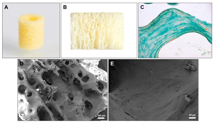
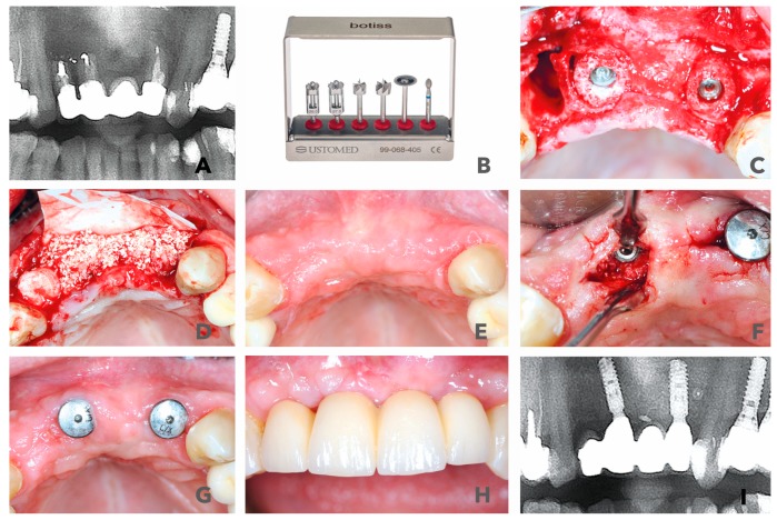
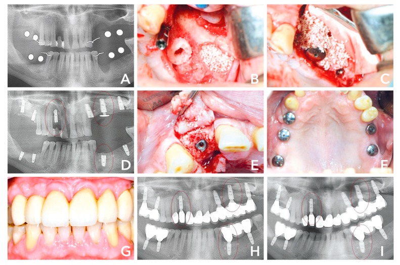
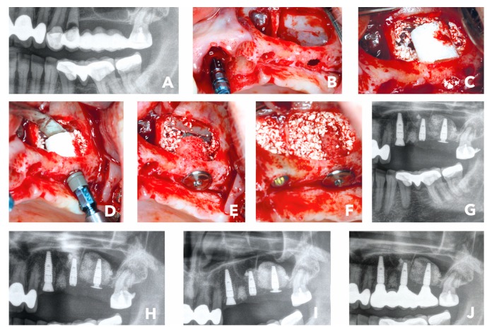
The main objective of this manuscript was to demonstrate the use of freeze-dried bone allografts (FDBA) by means of a technique of simultaneous bone augmentation and implant placement ("Bone Ring Technique") in different indications, i.e., ridge reconstruction and sinus floor elevation procedure with a maxillary bone height of less than 4 mm. Moreover, cases with an up to 3-year follow-up were chosen to analyze the techniques of mid-term clinical success. Finally, the purification success of the FDBA was analyzed by means of established scanning electron microscopic (SEM) and histological methods. The FBDA bone ring was applied in three different patients and indications and presented; the healing success was analyzed on the basis of radiographical and clinical images. For analysis of the purification of the allogeneic bone, previously established histological methods and scanning electron microscopy (SEM) were applied. All analyzed patient cases showed that the FDBA-based bone ring was fully integrated into newly built alveolar bone. Furthermore, the observations revealed that the three-dimensional bone reconstructions in maxilla and mandible were stable within the observational period of up to 3 years. Altogether, the present data show that the application of the Bone Ring Technique using the FDBA rings allows for successful regeneration of alveolar bone with a predictable clinical outcome, functionality and esthetics. Moreover, the material analyses showed that the allogeneic bone tissue was free of cells or cell remnants, while the (ultra-) structure of the bone matrix has been retained. Thus, the biological safety of the FDBA has been confirmed.

Keywords: alveolar bone loss; alveolar bone regeneration; bone ring technique; freeze-dried bone allograft (FDBA); purification; sinus augmentation freeze-dried bone allografts (FDBA).


Conflict of interest statement

The authors declare no conflict of interest.

Figures

Figure 1
Figure 1
(A) Image of the bone ring that shows its porous structure. (B) Profile of the bone ring showing the trabecular subarrangement. (C) Exemplary histological image of the former bone matrix indicating its lamellar architecture and its complete clearance from cells as well as the matrix-associated collagen fibers (arrow) (Masson Goldner staining, 400x magnification, scale bar = 20 µm). (D) Exemplary SEM image that shows the lamellar structure of the bone substitute material and the macro- and micro-porosity (40x magnfication). (E) SEM image of the material surface that shows the clearance of the osteocyte lacunae and the fibre-like surface pattern (200x magnification).
Figure 2
Figure 2
(A) Radiological display of the bone defect in frontal maxilla #11-22. (B) maxgraft bonering surgical kit. (C) Bone augmentation with two cylindrical FDBAs and fixation with dental implants. (D) Augmentation site contoured with bovine bone substiute and covered with collagen membrane. (E) and (F) Six-month follow-up of contouring and re-entry to place gingiva formers. (G,H) Integration of prosthetic restoration 6 weeks after re-entry and 7.5 months after initial surgery. (I) Thirty-six-month radiological follow-up showed stable bone in augmented area.
Figure 3
Figure 3
(A) Initial situation 6 weeks after extraction of teeth #12, #24-25 and #34. (B) Second quadrant: implant placement #28, sinus floor elevation and bone augmenation with a 7-mm bone ring block and immediate implant placement #26. (C) Sinus cavity filled with bovine bone substiute and covered with a collagene membrane. (D) Radiological control 8 months after surgery with stable bone around the shouders of the implants. (E) After opening the flap, the graft seemed to be well integrated. (F) Five weeks after re-entry and 9 months after initial surgery healthy soft tissues was observable. (G) Prosthtetic restoration in place. (H) Radiological control 14 months after surgery, (I) compared to 36 months, showed hard tissue maturation and stable dental implants.
Figure 4
Figure 4
(A) Initial situation: Radiological image of bridge retained second quadrant with hopeless teeth #23 and #24. (B) External sinus floor elavation and implant placement #23. (C) Placement of a 7-mm bone ring block #27 and bovine bone substitute. (D) Implant placement. (E) Crestal placement of a fication screw to secure the bone graft and implant. (F) Implant placement in #25 and sinus cavity filled with bovine bone substitute. (G) Post-op x-ray. (H) Radiographical control 6 months after surgery. (I) Radiographical control 1 week later when the implants were uncovered and prosthodontic impression made. (J) Radiographical image of final prosthetic restoration 6.5 months after initial surgery.

References

    1. Buser D., Chappuis V., Belser U.C., Chen S. Implant placement post extraction in esthetic single tooth sites: When immediate, when early, when late? Periodontol 2000. 2017;73:84–102. doi: 10.1111/prd.12170. - DOI - PubMed
    1. Sheikh Z., Najeeb S., Khurshid Z., Verma V., Rashid H., Glogauer M. Biodegradable Materials for Bone Repair and Tissue Engineering Applications. Materials (Basel) 2015;8:5744–5794. doi: 10.3390/ma8095273. - DOI - PMC - PubMed
    1. Cicciu M., Cervino G., Herford A.S., Fama F., Bramanti E., Fiorillo L., Lauritano F., Sambataro S., Troiano G., Laino L. Facial Bone Reconstruction Using both Marine or Non-Marine Bone Substitutes: Evaluation of Current Outcomes in a Systematic Literature Review. Mar. Drugs. 2018;16:27. doi: 10.3390/md16010027. - DOI - PMC - PubMed
    1. Sakkas A., Wilde F., Heufelder M., Winter K., Schramm A. Autogenous bone grafts in oral implantology-is it still a “gold standard”? A consecutive review of 279 patients with 456 clinical procedures. Int. J. Implant Dent. 2017;3:23. doi: 10.1186/s40729-017-0084-4. - DOI - PMC - PubMed
    1. Nkenke E., Weisbach V., Winckler E., Kessler P., Schultze-Mosgau S., Wiltfang J., Neukam F.W. Morbidity of harvesting of bone grafts from the iliac crest for preprosthetic augmentation procedures: A prospective study. Int. J. Oral Maxillofac. Surg. 2004;33:157–163. doi: 10.1054/ijom.2003.0465. - DOI - PubMed

Publication types

LinkOut - more resources