. 2019 Apr 19;9(4):637.
doi: 10.3390/nano9040637.

Plasma-Coated Polycaprolactone Nanofibers with Covalently Bonded Platelet-Rich Plasma Enhance Adhesion and Growth of Human Fibroblasts

Affiliations

Plasma-Coated Polycaprolactone Nanofibers with Covalently Bonded Platelet-Rich Plasma Enhance Adhesion and Growth of Human Fibroblasts

Svetlana Miroshnichenko et al. Nanomaterials (Basel). .

Abstract

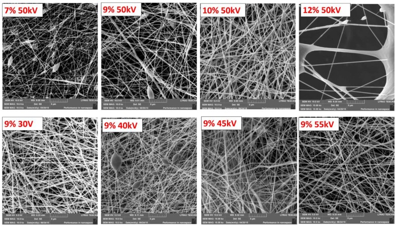
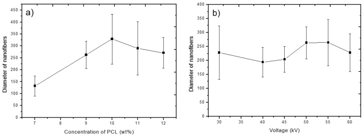
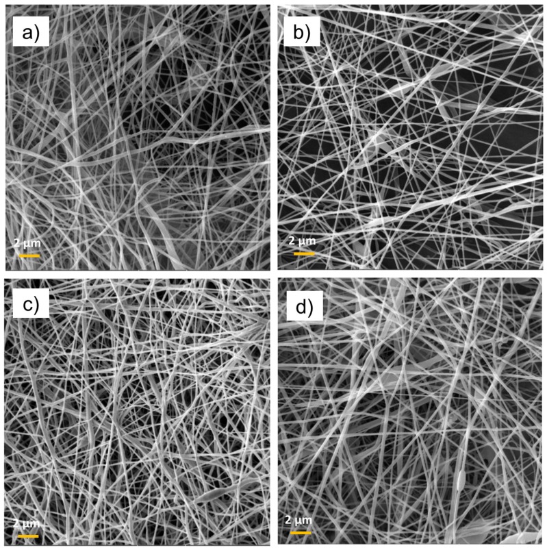
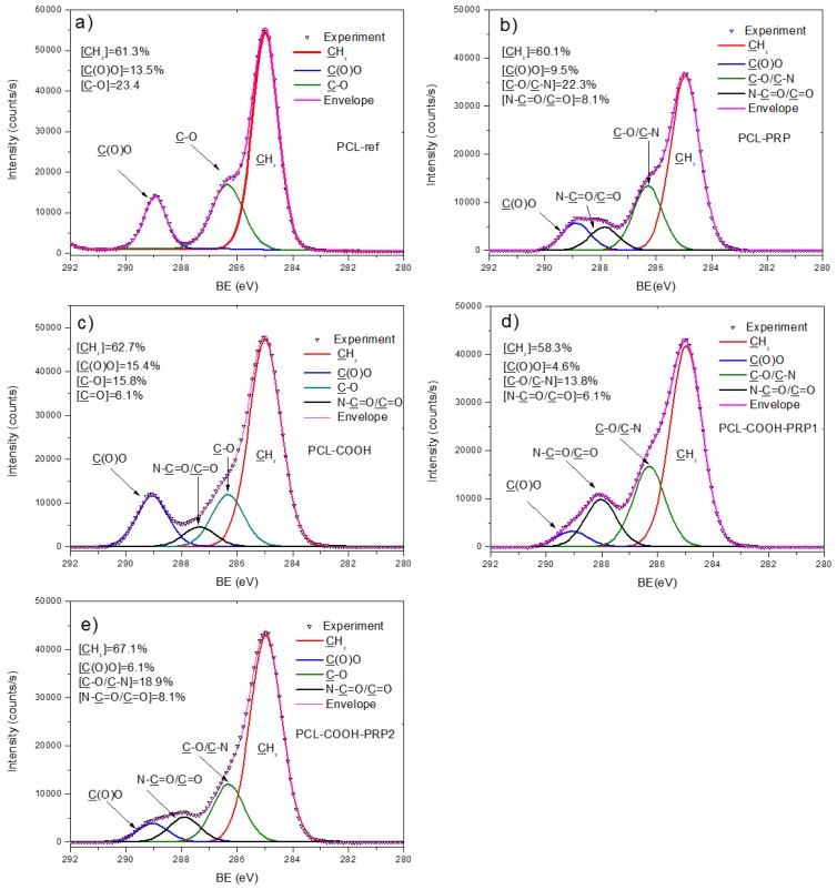
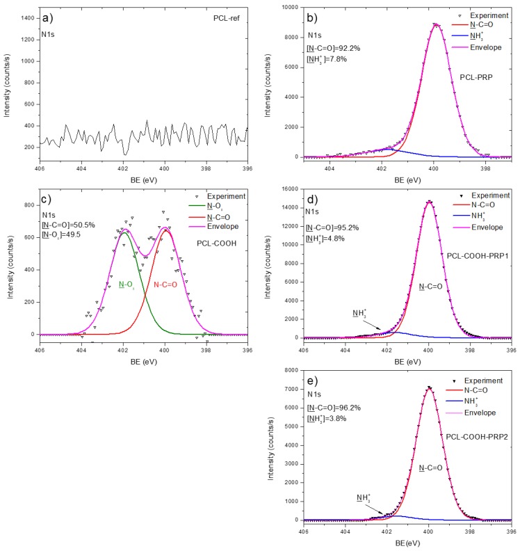
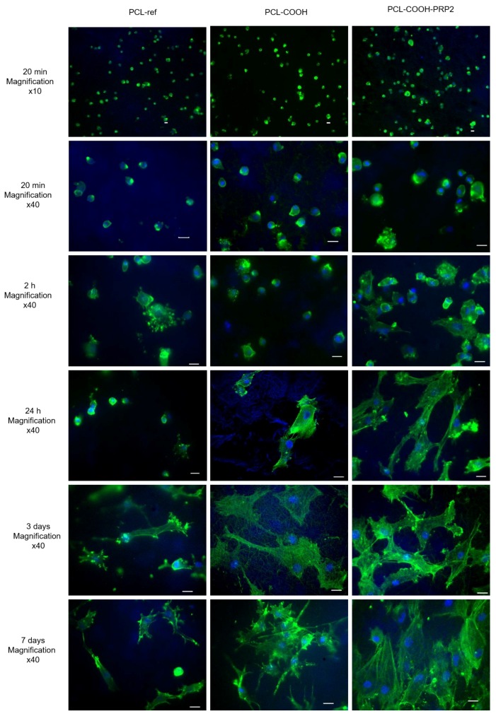
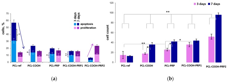
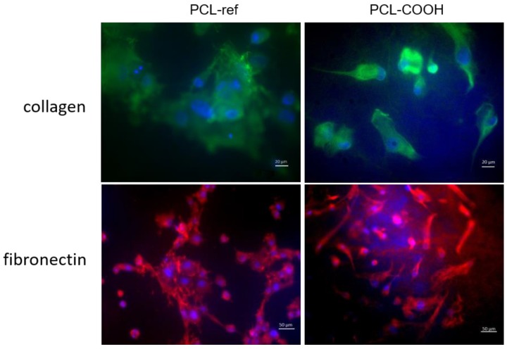
Biodegradable nanofibers are extensively employed in different areas of biology and medicine, particularly in tissue engineering. The electrospun polycaprolactone (PCL) nanofibers are attracting growing interest due to their good mechanical properties and a low-cost structure similar to the extracellular matrix. However, the unmodified PCL nanofibers exhibit an inert surface, hindering cell adhesion and negatively affecting their further fate. The employment of PCL nanofibrous scaffolds for wound healing requires a certain modification of the PCL surface. In this work, the morphology of PCL nanofibers is optimized by the careful tuning of electrospinning parameters. It is shown that the modification of the PCL nanofibers with the COOH plasma polymers and the subsequent binding of NH2 groups of protein molecules is a rather simple and technologically accessible procedure allowing the adhesion, early spreading, and growth of human fibroblasts to be boosted. The behavior of fibroblasts on the modified PCL surface was found to be very different when compared to the previously studied cultivation of mesenchymal stem cells on the PCL nanofibrous meshes. It is demonstrated by X-ray photoelectron spectroscopy (XPS) that the freeze-thawed platelet-rich plasma (PRP) immobilization can be performed via covalent and non-covalent bonding and that it does not affect biological activity. The covalently bound components of PRP considerably reduce the fibroblast apoptosis and increase the cell proliferation in comparison to the unmodified PCL nanofibers or the PCL nanofibers with non-covalent bonding of PRP. The reported research findings reveal the potential of PCL matrices for application in tissue engineering, while the plasma modification with COOH groups and their subsequent covalent binding with proteins expand this potential even further. The use of such matrices with covalently immobilized PRP for wound healing leads to prolonged biological activity of the immobilized molecules and protects these biomolecules from the aggressive media of the wound.

Keywords: COOH plasma; cell adhesion and spreading; cell viability; freeze–thawed platelet-rich plasma immobilization; nanofibers; polycaprolactone.


Conflict of interest statement

The authors declare no conflicts of interest.

Figures

Figure 1
Figure 1
SEM micrographs of the polycaprolactone (PCL-ref) nanofibers obtained from the PCL solutions with the different PCL concentrations and at different voltages. The PCL concentration and voltage are reported on the images. The size of the bar corresponds to 1 µm.
Figure 2
Figure 2
Thickness of resulting PCL nanofibers as a function of the PCL concentration (a) and applied voltage (b) during the electrospinning process. Error bars represent standard deviation of the nanofibers’ thickness distribution.
Figure 3
Figure 3
SEM micrographs of PCL-ref (a), PCL/platelet-rich plasma (PCL-PRP) (b), PCL-COOH (c), and PCL-COOH-PRP2 (d) The size of the bar corresponds to 1 µm.
Figure 4
Figure 4
X-ray photoelectron spectroscopy (XPS) C 1s curve fitting of PCL-ref (a), PCL-PRP (b), PCL-COOH (c), PCL-COOH-PRP1 (d), and PCL-COOH-PRP2 (e).
Figure 5
Figure 5
XPS N 1s curve fitting of PCL-ref (a), PCL-PRP (b), PCL-COOH (c), PCL-COOH-PRP1 (d), and PCL-COOH-PRP2 (e).
Figure 6
Figure 6
Fibroblast cell adhesion and spreading on the surface of PCL-ref, PCL-COOH, and PCL-COOH-PRP2 after 20 min, 24 h, and three and seven days of cultivation. The cytoskeleton actin filaments were stained by phalloidin (green), the cell nuclei were stained by Hoechst 33342 (blue). The size of the bar corresponds to 20 μm.
Figure 7
Figure 7
The influence of scaffold surface on (a) cell proliferation and apoptosis, and (b) cell count for three and seven days of cultivation. The level of proliferation rate was estimated by determining the ratio of total cells (Hoechst-positive) to the EdU-positive cells. The percentage of apoptotic cells was calculated as the ratio of the nuclei with chromatin condensation and nuclear fragmentation (kariorhexis) to nuclei with homogeneous coloration. The arrows indicate the relationship of the level of cell apoptosis and cell proliferation. Data are expressed as means ± standard errors; ** p < 0.01, * p < 0.05.
Figure 8
Figure 8
Fibronectin (FN) and collagen secretion by the fibroblasts seeded on PCL-ref and PCL-COOH. Cells were stained by antibody Alexa Fluor 594 (orange) to collagen (magnification 40×) and Alexa Fluor 488 to FN (magnification 20×).
Figure 9
Figure 9
Scheme of “electrostatic” and covalent immobilization of FN on PCL-COOH.

Similar articles

Cited by

References

    1. Lev-Tov H., Li C.-S., Dahle S., Isseroff R.R. Cellular versus acellular matrix devices in treatment of diabetic foot ulcers: Study protocol for a comparative efficacy randomized controlled trial. Trials. 2013;14:8. doi: 10.1186/1745-6215-14-8. - DOI - PMC - PubMed
    1. Watt S.M., Pleat J.M. Stem cells, niches and scaffolds: Applications to burns and wound care. Adv. Drug Deliv. Rev. 2018;123:82–106. doi: 10.1016/j.addr.2017.10.012. - DOI - PubMed
    1. Dickinson L.E., Gerecht S. Engineered Biopolymeric Scaffolds for Chronic Wound Healing. Front. Physiol. 2016;7:623. doi: 10.3389/fphys.2016.00341. - DOI - PMC - PubMed
    1. Chaudhari A.A., Vig K., Baganizi D.R., Sahu R., Dixit S., Dennis V., Singh S.R., Pillai S.R., Hardy J.G. Future Prospects for Scaffolding Methods and Biomaterials in Skin Tissue Engineering: A Review. Int. J. Mol. Sci. 2016;17:1974. doi: 10.3390/ijms17121974. - DOI - PMC - PubMed
    1. Manakhov A., Kedroňová E., Medalová J., Černochová P., Obrusník A., Michlíček M., Shtansky D.V., Zajíčková L. Carboxyl-anhydride and amine plasma coating of PCL nanofibers to improve their bioactivity. Mater. Des. 2017;132:257–265. doi: 10.1016/j.matdes.2017.06.057. - DOI