. 2019 Jul 15;25(14):4400-4412.
doi: 10.1158/1078-0432.CCR-18-3183. Epub 2019 Apr 22.

Engineering the TGFβ Receptor to Enhance the Therapeutic Potential of Natural Killer Cells as an Immunotherapy for Neuroblastoma

Affiliations

Engineering the TGFβ Receptor to Enhance the Therapeutic Potential of Natural Killer Cells as an Immunotherapy for Neuroblastoma

Rachel A Burga et al. Clin Cancer Res. .

Abstract

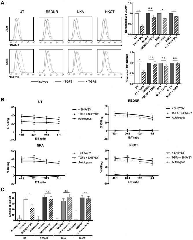
Purpose: The ability of natural killer (NK) cells to lyse allogeneic targets, without the need for explicit matching or priming, makes them an attractive platform for cell-based immunotherapy. Umbilical cord blood is a practical source for generating banks of such third-party NK cells for "off-the-shelf" cell therapy applications. NK cells are highly cytolytic, and their potent antitumor effects can be rapidly triggered by a lack of HLA expression on interacting target cells, as is the case for a majority of solid tumors, including neuroblastoma. Neuroblastoma is a leading cause of pediatric cancer-related deaths and an ideal candidate for NK-cell therapy. However, the antitumor efficacy of NK cells is limited by immunosuppressive cytokines in the tumor microenvironment, such as TGFβ, which impair NK cell function and survival.

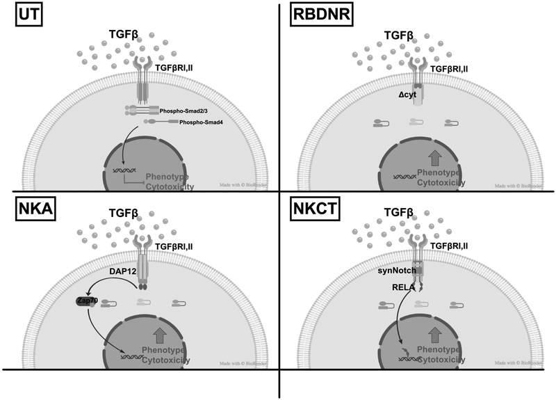
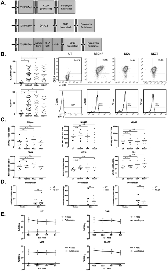
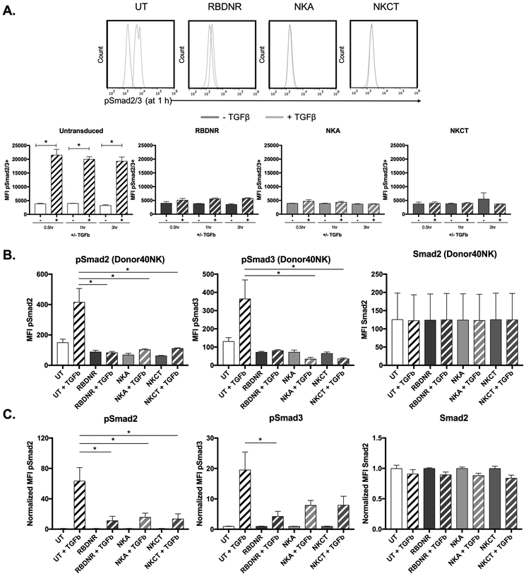
Experimental design: To overcome this, we genetically modified NK cells to express variant TGFβ receptors, which couple a mutant TGFβ dominant-negative receptor to NK-specific activating domains. We hypothesized that with these engineered receptors, inhibitory TGFβ signals are effectively converted to activating signals.

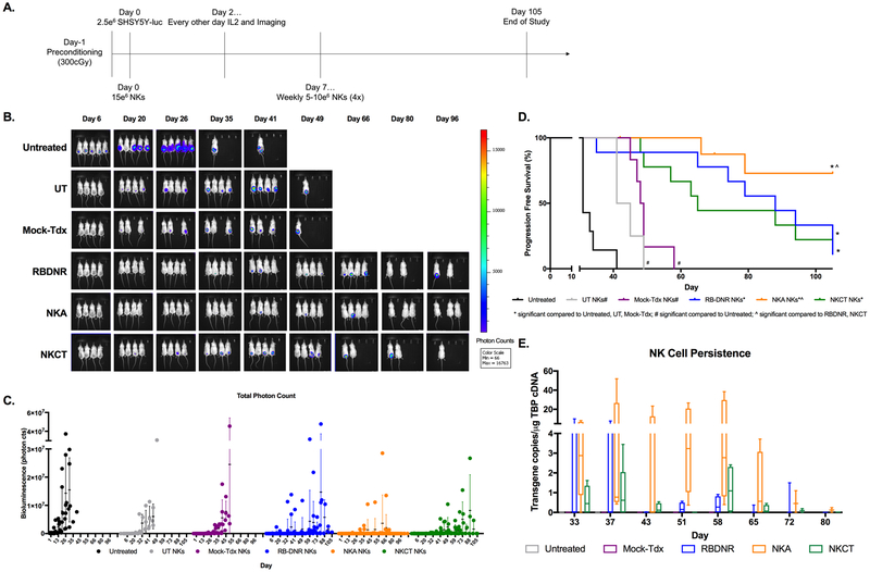
Results: Modified NK cells exhibited higher cytotoxic activity against neuroblastoma in a TGFβ-rich environment in vitro and superior progression-free survival in vivo, as compared with their unmodified controls.

Conclusions: Our results support the development of "off-the-shelf" gene-modified NK cells, that overcome TGFβ-mediated immune evasion, in patients with neuroblastoma and other TGFβ-secreting malignancies.


Conflict of interest statement

The authors declare no potential conflicts of interest.

Figures

Fig. 1.
Fig. 1.. TGFβ signaling in untransduced vs. RBDNR, NKA, or NKCT TGFβ-receptor modified NK cells.
Schematic depicting the effects of TGFβ binding to the receptor complex: Untransduced (UT) NK cells express the wild-type TGFβRII, which, when engaged with TGFβ in the tumor microenvironment, initiates a signaling cascade that culminates in impaired NK cell phenotype and cytotoxicity. NK cells transduced with the RBDNR, NKA, or NKCT variant TGFβ receptors alter the intracellular signaling and allow for maintained or enhanced NK cell phenotype and cytotoxicity in the setting of tumor-associated TGFβ.
Fig. 2.
Fig. 2.. Generating and characterizing TGFβ receptor-modified NK cells.
(A) Vector maps of RBDNR (top), NKA (middle), and NKCT (bottom) constructs. (B) Flow cytometry demonstrating transduction efficiency based on TGFβRII and/or CD19 positive staining. Representative flow dot plots and histograms are on the right, and summarizing data on the left. (C) The phenotype of transduced and untransduced NK cells were examined by flow cytometry, and mean fluorescent intensity values for a given surface receptor is depicted in each panel. (D) Transduced and untransduced NK cells were stained with CFSE, and stimulated with irradiated feeder cells. After 3 days, cells were harvested and assessed for CFSE dilution by flow cytometry. (E) 51Cr-labeled K562 target cells were co-cultured at various effector:target ratios with transduced or untransduced NK cells, and cytotoxicity after 5 hour co-culture was determined based on chromium content in the supernatant, calculated with spontaneous and maximum release controls. All data is representative of experiments with >8 donor lines, with * indicating significant p values <0.05.
Fig. 3.
Fig. 3.. Examining the molecular effects of TGFβ-signaling.
(A) Flow cytometry was performed to examine the expression of phosphorylated Smad2/3 in transduced and untransduced NK cells after 0.5, 1, and 3 h of exposure to 10 ng/mL TGFβ. Representative histograms are on top, and summarizing data below. (B) Protein was isolated from transduced and untransduced NK cells after 1 hour of exposure to 10 ng/mL TGFβ, and was assessed for phosphorylated Smad2, phosphorylated Smad3, and Smad2 protein content by multiplex assay. Representative protein data for NK cells generated from one donor line. (C) Summarizing protein data for NK cells, where protein amounts are normalized to that of non-TGFβ conditions. All data is representative of experiments with >3 donor lines, with * indicating significant p values <0.05.
Fig. 4.
Fig. 4.. Examining downstream phenotypic and functional effects of TGFβ-signaling.
(A) Transduced and untransduced NK cells were exposed to TGFβ for 5 days, after which they were harvested and examined for phenotypic changes by flow cytometry. Representative histograms on the left and summarizing data on the right demonstrates changes in the expression of DNAM1 and NKG2D, with mean fluorescent intensities normalized to that of non-TGFβ conditions. (B) 51Cr-labeled SHSY5Y neuroblastoma cells were co-cultured at various effector:target ratios with transduced or untransduced NK cells, and cytotoxicity after 5 hour co-culture was determined based on chromium content in the supernatant, calculated with spontaneous and maximum release controls. (C) Cytotoxicity of NK cells against SHSY5Y neuroblastoma at a 40:1 effector:target ratio. All data is representative of experiments with >7 donor lines, with * indicating significant p values <0.05.
Fig. 5.
Fig. 5.. Evaluation of TGFβ-signaling induced NK cell activation.
(A) Flow cytometry was performed to examine the expression of p65 (RELA) in transduced and untransduced NK cells after 0.5, 1, and 3 h of exposure to 10 ng/mL TGFβ. (B) Representative histograms for flow cytometry of p65(RELA) expression on untransduced, RBDNR, NKA, and NKCT NK cells following 1 h of exposure to 10 ng/mL TGFβ. (C) Protein was isolated from transduced and untransduced NK cells after 1 hour of exposure to 10 ng/mL TGFβ, and was assessed for phosphorylated ERK1/2 and phosphorylated Akt protein content by multiplex assay. (D) Supernatant was isolated from NK cell cultures after 12 h of exposure to 10 ng/mL TGFβ and concentration of TNFa and IFNy was quantified by multiplex assay. Summarizing protein and cytokine data is graphed, where protein amounts are normalized to that of non-TGFβ conditions. All data is representative of experiments with >3 donor lines, with * indicating significant p values <0.05.
Fig. 6.
Fig. 6.. Long-term tumor-free survival with repeat doses of NK cell treatment in vivo.
(A) Schematic for our in vivo neuroblastoma model: immunodeficient mice were preconditioned, inoculated with luciferase-positive SHSY5Y, treated with systemically delivered transduced or untransduced NK cells on a weekly basis for 5 weeks, and received adjuvant IL2. (B) Tumor growth was monitored by evaluation bioluminescence of animals, which was (C) quantified by total photon counts taken at the same scale. (D) The effect of treatment with transduced or untransduced NK cells on animal survival over the length of the study. (E) Untransduced or transduced NK cells were identified using ddPCR methods to identify transgene copies in systemic blood isolated at weekly intervals following the last NK treatment. Tumor bioluminescence was qualitatively identified according to the heat map color scale, in vivo results are representative with n=5–9 animals/experimental group, ^ indicates significant p values <0.05 compared to RBDNR and NKCT animals, * indicates significant p values <0.05 compared to untreated, UT and Mock-tdx animals, and # indicates significant p values <0.05 compared to untreated animals only.

References

    1. Rosenberg SA, Restifo NP, Adoptive cell transfer as personalized immunotherapy for human cancer. Science 348, 62–68 (2015). - PMC - PubMed
    1. Restifo NP, Dudley ME, Rosenberg SA, Adoptive immunotherapy for cancer: harnessing the T cell response. Nat Rev Immunol 12, 269–281 (2012). - PMC - PubMed
    1. Tey SK, Bollard CM, Heslop HE, Adoptive T-cell transfer in cancer immunotherapy. Immunol Cell Biol 84, 281–289 (2006). - PubMed
    1. Kalos M, June CH, Adoptive T cell transfer for cancer immunotherapy in the era of synthetic biology. Immunity 39, 49–60 (2013). - PMC - PubMed
    1. Herberman RB, Nunn ME, Holden HT, Lavrin DH, Natural cytotoxic reactivity of mouse lymphoid cells against syngeneic and allogeneic tumors. II. Characterization of effector cells. Int J Cancer 16, 230–239 (1975). - PubMed

Publication types

MeSH terms

Substances