. 2019 Aug 22;201(18):e00064-19.
doi: 10.1128/JB.00064-19. Print 2019 Sep 15.

Role of Caulobacter Cell Surface Structures in Colonization of the Air-Liquid Interface

Affiliations

Role of Caulobacter Cell Surface Structures in Colonization of the Air-Liquid Interface

Aretha Fiebig. J Bacteriol. .

Abstract

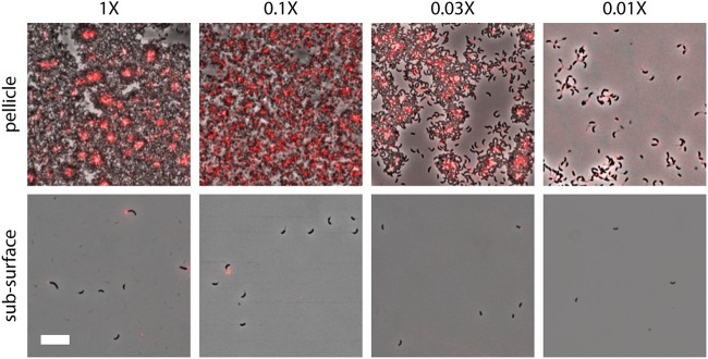
In aquatic environments, Caulobacter spp. can be found at the boundary between liquid and air known as the neuston. I report an approach to study temporal features of Caulobacter crescentus colonization and pellicle biofilm development at the air-liquid interface and have defined the role of cell surface structures in this process. At this interface, C. crescentus initially forms a monolayer of cells bearing a surface adhesin known as the holdfast. When excised from the liquid surface, this monolayer strongly adheres to glass. The monolayer subsequently develops into a three-dimensional structure that is highly enriched in clusters of stalked cells known as rosettes. As this pellicle film matures, it becomes more cohesive and less adherent to a glass surface. A mutant strain lacking a flagellum does not efficiently reach the surface, and strains lacking type IV pili exhibit defects in organization of the three-dimensional pellicle. Strains unable to synthesize the holdfast fail to accumulate at the boundary between air and liquid and do not form a pellicle. Phase-contrast images support a model whereby the holdfast functions to trap C. crescentus cells at the air-liquid boundary. Unlike the holdfast, neither the flagellum nor type IV pili are required for C. crescentus to partition to the air-liquid interface. While it is well established that the holdfast enables adherence to solid surfaces, this study provides evidence that the holdfast has physicochemical properties that allow partitioning of nonmotile mother cells to the air-liquid interface and facilitate colonization of this microenvironment.IMPORTANCE In aquatic environments, the boundary at the air interface is often highly enriched with nutrients and oxygen. Colonization of this niche likely confers a significant fitness advantage in many cases. This study provides evidence that the cell surface adhesin known as a holdfast enables Caulobacter crescentus to partition to and colonize the air-liquid interface. Additional surface structures, including the flagellum and type IV pili, are important determinants of colonization and biofilm formation at this boundary. Considering that holdfast-like adhesins are broadly conserved in Caulobacter spp. and other members of the diverse class Alphaproteobacteria, these surface structures may function broadly to facilitate colonization of air-liquid boundaries in a range of ecological contexts, including freshwater, marine, and soil ecosystems.

Keywords: Alphaproteobacteria; Caulobacter; biofilm; flagellum; holdfast; neuston; pellicle; type 4 pilus; unipolar polysaccharide.


Figures

FIG 1
FIG 1
Caulobacter crescentus strain CB15 develops a pellicle at the air-liquid interface during static growth. (A) Wild-type C. crescentus CB15 culture grown at room temperature without mixing (i.e., static growth) for 3 days. Note the accumulation of cells in a pellicle at the air-liquid interface at the top of the beaker. (B) Pellicle development requires growth. (Top) A culture was grown to stationary phase under aerated conditions, transferred to a fresh tube (far left), and serially diluted with fresh medium (toward the right; dilution fractions are shown above each tube). (Middle) The same tubes are shown after incubation on the benchtop for 4 days. The arrowhead highlights colonization of the air-liquid interface in diluted cultures that grew postdilution but not in the undiluted culture. (Bottom) Crystal violet (CV) stain of cells attached to the tubes after cultures were washed away. The arrowhead highlights the position of the air-liquid interface. (C) The oxygen gradient is steep at the surfaces of unmixed cultures. Oxygen concentration as a function of depth from the surface (0 mm) was measured in beakers in which PYE medium was left sterile or inoculated with wild-type C. crescentus CB15 or wild-type C. crescentus NA1000 and incubated without mixing. Each trace represents an independent culture (n = 2). The limit of detection is 0.3 μM. (D) Method for sampling the pellicle. The large end of a sterile pipet tip is touched on the pellicle surface (i), lifted (ii, iii), and placed on a glass slide (iv, v). A pellicle scar (vi, circled) can be seen after the plug was removed from this 72-h culture. (E) Pellicle plugs were placed on glass slides (left column) and then covered with a coverslip (middle column). Outlines of the plugs under coverslips are on the right. The heavy line corresponds to the edges of the plugs. Stationary bubbles that formed upon placement of the coverslip are filled in blue. The time since inoculation is indicated for each sample.
FIG 2
FIG 2
The pellicle develops from a homogeneous monolayer into a multilayered structure of dense rosettes. Surface plugs from a wild-type culture sampled periodically throughout static growth (the time after inoculation appears on the left) were evaluated by phase-contrast microscopy (left) and crystal violet (CV) staining (right). Two microscopy images are presented for each time point to capture the structure of cells in the center of the plug (left column) and also at the edges of the plug (right column; 8- to 36-h samples); cells disrupted from the multilayered plug structure were also imaged (right column; 48- to 96-h samples). Plug edges are outlined in Fig. 1E. White scale bar, 20 μm; black scale bar, 1 cm. This time course was repeated at least three times. Representative images from one experiment are presented.
FIG 3
FIG 3
In situ fWGA-stained pellicle samples. Phase-contrast and fluorescence images of cells grown in the presence of 1 μg/ml fWGA sampled at time intervals after inoculation. During the transition from a monolayer to a multilayer structure, at 32 h, two focal planes of the same position in the pellicle plug are presented. These images correspond to the uppermost plane, where fWGA bound to individual cells is in focus, and the bottom plane just below the monolayer, where the centers of rosettes are in focus. At 40 and 48 h, focal planes from the middle of the film are shown. Scale bar, 20 μm. Representative images from one of several time courses are presented.
FIG 4
FIG 4
Linear arrays of rosettes in the center of an excised pellicle plug. (A) In situ fWGA-stained surface film harvested 40 h after inoculation. The focal plane is just below a monolayer. Overlay (top), fWGA-stained holdfast (middle) and phase-contrast (bottom) images from one field of view (137 μm by 104 μm) are shown. Note, some of the rosette chains extend beyond the focal plane. (B) Crops corresponding to the regions boxed in panel A. Scale bars, 10 μm.
FIG 5
FIG 5
C. crescentus cells localized to the boundaries of air bubbles. Phase-contrast (left), fWGA-stained holdfast (middle) and overlay (right) micrographs from static cultures grown with fWGA, 8 to 12 h after inoculation. The interface between the air bubbles and the liquid medium is bright in phase-contrast images. The air and liquid sides of the boundary are indicated. Scale bar, 10 μm.
FIG 6
FIG 6
Macroscopic pellicles of polar appendage mutants. Static cultures of wild-type (CB15) and mutant strains 48 and 72 h after inoculation imaged from above and below, respectively. See the text for details on mutants. This experiment was repeated multiple times. The results of one representative experiment are shown.
FIG 7
FIG 7
Crystal violet staining of pellicle plug samples. Pellicles of wild-type (CB15) and mutant strains sampled throughout development were evaluated by crystal violet staining. Note the three stages of pellicle development (CB15 times are indicated): adhesive monolayer (up to 24 h), crumbly transition phase (32 to 40 h), and nonadhesive film (48+ hours). For each genotype, the beginning of the crumbly phase is marked with an asterisk. Pellicles sampled are from the same experiment whose results are presented in Fig. 8 This experiment was repeated two additional times. Scale bar, 1 cm.
FIG 8
FIG 8
C. crescentus mutants lacking polar appendages exhibit defects in pellicle development. Phase-contrast micrographs of pellicle samples from wild-type (CB15) and mutant strains were taken at 8-h intervals. Scale bar, 20 μm. Representative images from one of three independent experiments are shown.
FIG 9
FIG 9
Pellicle structures of NA1000 strains differ from those of CB15. (A) Pellicles of cultures grown statically for 3 days are pictured (top). After growth, the tubes were stained with crystal violet to highlight cells adhered to the glass at the surfaces of the cultures (bottom). Genotypes of strains are indicated above the tubes. Strains that differ only at the hfsA locus are paired, and the hfsA allele is indicated as the functional CB15 allele (+), the null allele (Δ), or the frameshifted NA1000 allele (fs). See the text for details about the mobile genetic element (MGE). A tube with sterile medium (left) demonstrates the characteristics of an uncolonized meniscus, similar to what is seen at the surface of medium colonized with strains lacking a functional hfsA allele. The whole surface of the culture is opaque when a pellicle film is present. WT, wild type. (B) Phase-contrast micrographs of pellicle samples from CB15, NA1000 hfsA+, and NA1000 ΔMGE hfsA+ pellicles collected 48 h after inoculation. The center of the plug (top) and rosettes disrupted from the film (bottom) were imaged. Scale bar, 20 μm.
FIG 10
FIG 10
Cells partition to the air-liquid boundary in dilute complex medium. Surface and subsurface samples of C. crescentus CB15 were cultured in increasingly dilute PYE medium. Dilution factors from the standard lab recipe (see Materials and Methods) are indicated at the top. Cultures were grown statically with fWGA to stain the holdfast for 2.5 days. Samples were imaged using phase-contrast and fluorescence imaging, and an overlay is presented for each condition. Scale bar, 10 μm.

Similar articles

Cited by

References

    1. Davies JT, Rideal EK. 1963. Interfacial phenomena, 2nd ed Academic Press, London, United Kingdom.
    1. Van Oss CJ. 2006. Interfacial forces in aqueous media, 2nd ed, p 438 Taylor & Francis, Boca Raton, FL.
    1. Marshall KC. 1996. Adhesion as a strategy for access to nutrients, p 59–87. In Fletcher M (ed), Bacterial adhesion: molecular and ecological diversity. Wiley-Liss, Inc, New York, NY.
    1. Henrici AT. 1933. Studies of freshwater bacteria: I. A direct microscopic technique. J Bacteriol 25:277–287. - PMC - PubMed
    1. Zobell CE, Allen EC. 1935. The significance of marine bacteria in the fouling of submerged surfaces. J Bacteriol 29:239–251. - PMC - PubMed

Publication types

MeSH terms

Substances