The Differential Expression of Mitochondrial Function-Associated Proteins and Antioxidant Enzymes during Bovine Herpesvirus 1 Infection: A Potential Mechanism for Virus Infection-Induced Oxidative Mitochondrial Dysfunction
- PMID: 31011285
- PMCID: PMC6442485
- DOI: 10.1155/2019/7072917
The Differential Expression of Mitochondrial Function-Associated Proteins and Antioxidant Enzymes during Bovine Herpesvirus 1 Infection: A Potential Mechanism for Virus Infection-Induced Oxidative Mitochondrial Dysfunction
Abstract
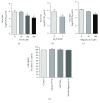
Reactive oxidative species (ROS) are important inflammatory mediators. Electrons escaping from the mitochondrial electron transport chain (ETC) during oxidative phosphorylation (OXPHOS) in the mitochondrial respiratory chain (RC) complexes contribute to ROS production. The cellular antioxidant enzymes are important for maintaining ROS release at the physiological levels. It has been reported that BoHV-1 infection induces overproduction of ROS and oxidative mitochondrial dysfunction in cell cultures. In this study, we found that chemical interruption of RC complexes by TTFA (an inhibitor of RC complex II), NaN3 (an inhibitor of RC complex IV), and oligomycin A (an inhibitor of ATP synthase) consistently decreased virus productive infection, suggesting that the integral processes of RC complexes are important for the virus replication. The virus infection significantly increased the expression of subunit SDHB (succinate dehydrogenase) and MTCO1 (cytochrome c oxidase subunit I), critical components of RC complexes II and IV, respectively. The expression of antioxidant enzymes including superoxide dismutase 1 (SOD1), SOD2, catalase (CAT), and glutathione peroxidase 4 (GPX4) was differentially affected following the virus infection. The protein TFAM (transcription factor A, mitochondrial) stimulated by either nuclear respiratory factor 1 (NRF1) or NRF2 is a key regulator of mitochondrial biogenesis. Interestingly, the virus infection at the late stage (at 16 h after infection) stimulated TFAM expression but decreased the levels of both NRF1 and NRF2, indicating that virus infection activated TFAM signaling independent of either NRF1 or NRF2. Overall, this study provided evidence that BoHV-1 infection altered the expression of molecules associated with RC complexes, antioxidant enzymes, and mitochondrial biogenesis-related signaling NRF1/NRF2/TFAM, which correlated with the previous report that virus infection induces ROS overproduction and mitochondrial dysfunction.
Figures


Similar articles
-
The neurogenic basic helix-loop-helix transcription factor NeuroD6 confers tolerance to oxidative stress by triggering an antioxidant response and sustaining the mitochondrial biomass.ASN Neuro. 2010 May 24;2(2):e00034. doi: 10.1042/AN20100005. ASN Neuro. 2010. PMID: 20517466 Free PMC article.
-
Sodium azide induces mitochondria‑mediated apoptosis in PC12 cells through Pgc‑1α‑associated signaling pathway.Mol Med Rep. 2019 Mar;19(3):2211-2219. doi: 10.3892/mmr.2019.9853. Epub 2019 Jan 14. Mol Med Rep. 2019. PMID: 30664159 Free PMC article.
-
Biochemical and transcriptional analyses of cadmium-induced mitochondrial dysfunction and oxidative stress in human osteoblasts.J Toxicol Environ Health A. 2018;81(15):705-717. doi: 10.1080/15287394.2018.1485122. Epub 2018 Jun 18. J Toxicol Environ Health A. 2018. PMID: 29913117
-
Regulatory crosstalk between the oxidative stress-related transcription factor Nfe2l2/Nrf2 and mitochondria.Toxicol Appl Pharmacol. 2018 Nov 15;359:24-33. doi: 10.1016/j.taap.2018.09.014. Epub 2018 Sep 18. Toxicol Appl Pharmacol. 2018. PMID: 30236989 Review.
-
Oxidative stress and mitochondrial dysfunction in sepsis: a potential therapy with mitochondria-targeted antioxidants.Infect Disord Drug Targets. 2009 Aug;9(4):376-89. doi: 10.2174/187152609788922519. Infect Disord Drug Targets. 2009. PMID: 19689380 Review.
Cited by
-
Effect of Melatonin on Herpesvirus Type 1 Replication.Int J Mol Sci. 2024 Apr 4;25(7):4037. doi: 10.3390/ijms25074037. Int J Mol Sci. 2024. PMID: 38612846 Free PMC article.
-
Carnitine palmitoyl-transferase 1A is potentially involved in bovine herpesvirus 1 productive infection.Vet Microbiol. 2024 Jan;288:109932. doi: 10.1016/j.vetmic.2023.109932. Epub 2023 Nov 30. Vet Microbiol. 2024. PMID: 38043447 Free PMC article.
-
Glycyrrhizin alleviates BoAHV-1-induced lung injury in guinea pigs by inhibiting the NF-κB/NLRP3 Signaling pathway and activating the Nrf2/HO-1 Signaling pathway.Vet Res Commun. 2024 Aug;48(4):2499-2511. doi: 10.1007/s11259-024-10436-7. Epub 2024 Jun 12. Vet Res Commun. 2024. PMID: 38865040
-
Integrated analysis of microRNA and messenger RNA expression profiles reveals functional microRNA in infectious bovine rhinotracheitis virus-induced mitochondrial damage in Madin-Darby bovine kidney cells.BMC Genomics. 2024 Feb 8;25(1):158. doi: 10.1186/s12864-024-10042-6. BMC Genomics. 2024. PMID: 38331736 Free PMC article.
-
NFAT5 Restricts Bovine Herpesvirus 1 Productive Infection in MDBK Cell Cultures.Microbiol Spectr. 2023 Aug 17;11(4):e0011723. doi: 10.1128/spectrum.00117-23. Epub 2023 May 25. Microbiol Spectr. 2023. PMID: 37227295 Free PMC article.
References
MeSH terms
Substances
LinkOut - more resources
Full Text Sources
Medical
Miscellaneous

