

Transorbital penetrating head trauma leading to serious cerebral edema - A case report
- PMID: 31011615
- PMCID: PMC6463219
- DOI: 10.1016/j.tcr.2019.100192
Transorbital penetrating head trauma leading to serious cerebral edema - A case report
Abstract
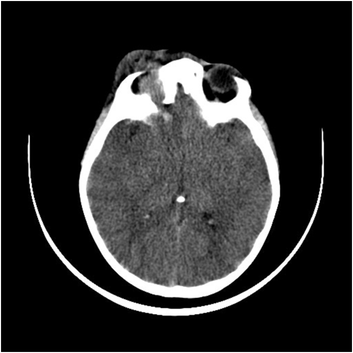
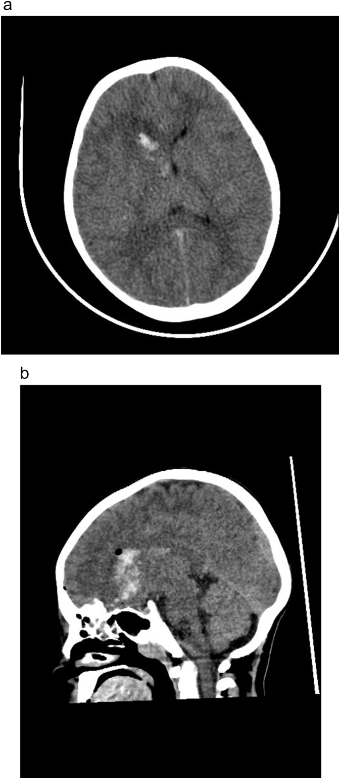
Penetrating transorbital head injuries are rarely seen. We present a 6-year old male patient who was referred to our department after a wooden stick penetrated his right eye. On admission his general condition was well and he showed no neurologic deficits. On his cranial computed tomography (CT) there was a fracture on his right orbital wall and minimal subarachnoid hemorrhage in his right frontal lobe. On later follow-ups the patient's condition worsened and his control CT showed diffuse cerebral edema. The patient underwent emergent decompressive surgery. Due to this immediate intervention the patient was released from the hospital with no major deficits later. Up to our knowledge, there are no cases in the literature reported in which decompressive craniectomy was necessary after a transorbital penetrating head trauma.
Figures


References
-
- Jandial R., Reichwage B., Levy M., Duenas V., Sturdivan L. Ballistics for the neurosurgeon. Neurosurgery. 2008;62(2):472–480. - PubMed
-
- Liu W.H., Chiang Y.H., Hsieh C.T., Sun J.M., Hsia C.C. Transorbital penetrating brain injury by branchlet: a rare case. J. Emerg. Med. 2011;41(5):482–485. - PubMed
-
- Fujimoto S., Onuma T., Amagasa M., Okudaira Y. Three cases of an intracranial wooden foreign body. No Shinkei Geka. 1987;15(7):751–756. - PubMed
-
- Kazarian E.L., Stokes N.A., Flyn J.T. The orbital puncture wound. Intracranial complications of a retained foreign body. J. Pediatr. Ophthalmol. Strabismus. 1980;17(4):247–250. - PubMed
Publication types
LinkOut - more resources
Full Text Sources
