

White matter hyperintensities in vascular contributions to cognitive impairment and dementia (VCID): Knowledge gaps and opportunities
- PMID: 31011621
- PMCID: PMC6461571
- DOI: 10.1016/j.trci.2019.02.001
White matter hyperintensities in vascular contributions to cognitive impairment and dementia (VCID): Knowledge gaps and opportunities
Abstract
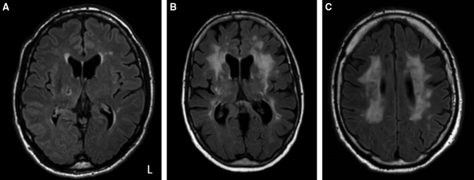
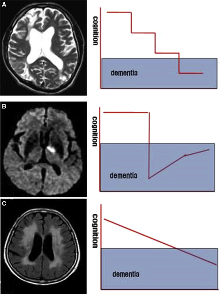
White matter hyperintensities (WMHs) are frequently seen on brain magnetic resonance imaging scans of older people. Usually interpreted clinically as a surrogate for cerebral small vessel disease, WMHs are associated with increased likelihood of cognitive impairment and dementia (including Alzheimer's disease [AD]). WMHs are also seen in cognitively healthy people. In this collaboration of academic, clinical, and pharmaceutical industry perspectives, we identify outstanding questions about WMHs and their relation to cognition, dementia, and AD. What molecular and cellular changes underlie WMHs? What are the neuropathological correlates of WMHs? To what extent are demyelination and inflammation present? Is it helpful to subdivide into periventricular and subcortical WMHs? What do WMHs signify in people diagnosed with AD? What are the risk factors for developing WMHs? What preventive and therapeutic strategies target WMHs? Answering these questions will improve prevention and treatment of WMHs and dementia.
Keywords: Leukoaraiosis; Small vessel disease; Vascular cognitive impairment; Vascular dementia; White matter lesions.
Figures
References
-
- Prins N.D., Scheltens P. White matter hyperintensities, cognitive impairment and dementia: an update. Nat Rev Neurol. 2015;11:157–165. - PubMed
-
- Kloppenborg R.P., Nederkoorn P.J., Geerlings M.I., van den Berg E. Presence and progression of white matter hyperintensities and cognition: a meta-analysis. Neurology. 2014;82:2127–2138. - PubMed
Publication types
Grants and funding
- P30 AG010124/AG/NIA NIH HHS/United States
- RF1 AG059421/AG/NIA NIH HHS/United States
- G0800701/1/NC3RS_/National Centre for the Replacement, Refinement and Reduction of Animals in Research/United Kingdom
- U54 HD083211/HD/NICHD NIH HHS/United States
- F31 AG054084/AG/NIA NIH HHS/United States
- R01 NS104127/NS/NINDS NIH HHS/United States
- MR/L023784/2/MRC_/Medical Research Council/United Kingdom
- P30 AG028383/AG/NIA NIH HHS/United States
- P50 AG033514/AG/NIA NIH HHS/United States
- F30 AG054115/AG/NIA NIH HHS/United States
- R01 NS017950/NS/NINDS NIH HHS/United States
- R01 AG054076/AG/NIA NIH HHS/United States
- MR/R005567/1/MRC_/Medical Research Council/United Kingdom
- F32 AG058395/AG/NIA NIH HHS/United States
- T32 GM008692/GM/NIGMS NIH HHS/United States
- R01 AG062572/AG/NIA NIH HHS/United States
- P30 AG049638/AG/NIA NIH HHS/United States
- P30 AG062715/AG/NIA NIH HHS/United States
- P30 AG010129/AG/NIA NIH HHS/United States
LinkOut - more resources
Full Text Sources
Other Literature Sources
Medical