. 2019 May;52(3):e12610.
doi: 10.1111/cpr.12610. Epub 2019 Apr 22.

Circular RNA cMras inhibits lung adenocarcinoma progression via modulating miR-567/PTPRG regulatory pathway

Affiliations

Circular RNA cMras inhibits lung adenocarcinoma progression via modulating miR-567/PTPRG regulatory pathway

Chengtao Yu et al. Cell Prolif. 2019 May.

Abstract

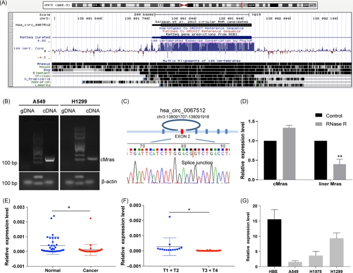
Objectives: Circular RNA, a type of RNA formed by a covalently closed loop, possesses sophisticated abilities of gene regulation in tumorigenesis and metastasis. However, the role of circRNAs on lung adenocarcinoma (LUAD) remains largely unknown.

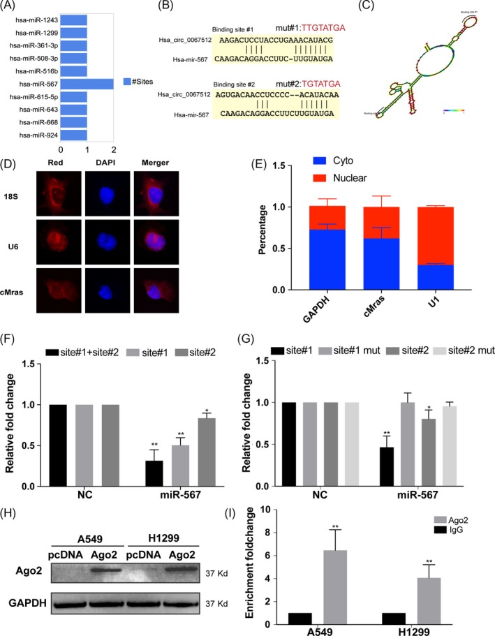
Materials and methods: The role of cMras was examined both in vitro and in vivo. cMras expression in LUAD tissues was determined by quantitative real-time PCR (qRT-PCR). Downstream targets of cMras were predicted by bioinformatics tools and confirmed by RNA immunoprecipitation assay and luciferase assay. qRT-PCR and western blot assay were used to detect the expression of specific targets.

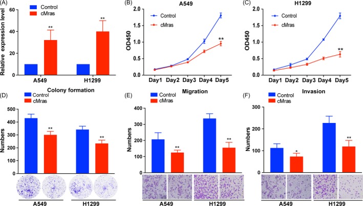
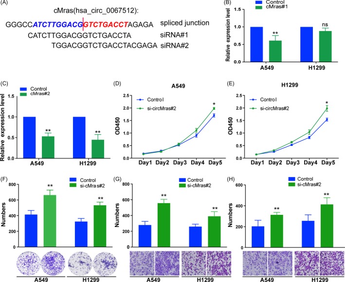
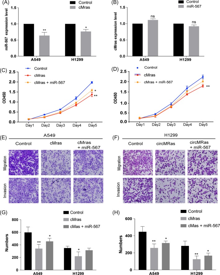
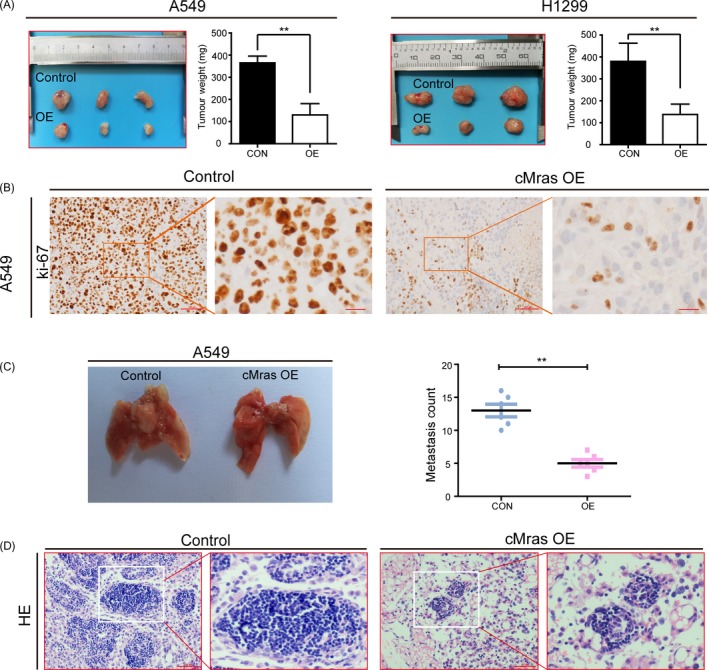
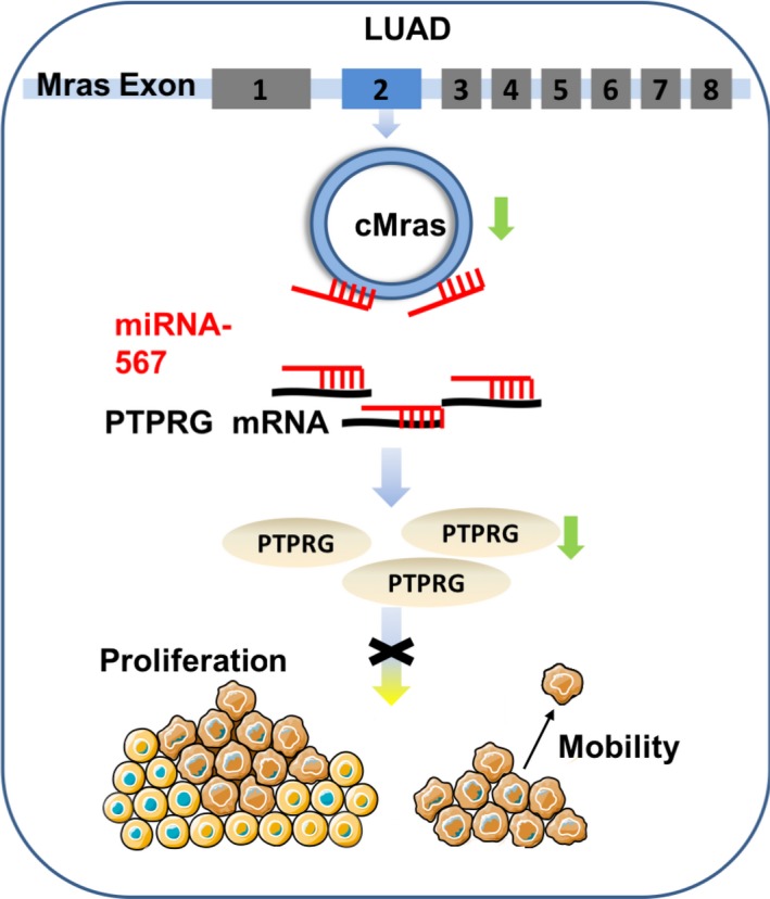
Results: Thirty-six paired LUAD and healthy tissues were collected and cMras resulted significantly downregulated in cancerous tissues. Its expression was negatively associated with tumour stages. cMras overexpression suppressed LUAD growth and metastasis, while endogenous cMras silencing resulted in the opposite effects. Bioinformatics analysis and experimental evidence confirmed that cMras was a sponge of miRNA-567 and released its direct target, PTPRG. cMras overexpression decreased miR-567 expression and subsequently increased PTPRG expression, while increased miRNA-567 expression blocked the effects induced by cMras. Moreover, PTPRG was downregulated in LUAD and patients with low PTPRG expression exhibited significantly poor prognosis. These results suggested that cMras/miR-567/PTPRG regulatory pathway might be associated to LUAD tumorigenesis and development.

Conclusions: A novel circular RNA cMras and its functions were identified, discovering a cMras/miR-567/PTPRG regulatory pathway in LUAD tumorigenesis and development.

Keywords: PTPRG; cMras; circular RNA; lung cancer; miR-567.


Conflict of interest statement

The authors declare no conflicts of interest.

Figures

Figure 1
Figure 1
cMras characterization and downregulation in LUAD. A, cMras chromosome location and species conservation were displayed by UCSC websites. B, Gel electrophoresis of cMras PCR products amplified by divergent primers in gDNA or cDNA. C, Sanger sequencing displayed back‐spliced junction of cMras, red line pointed back‐spliced site. D, cMras and linear Mras expression were detected by qRT‐PCR after RNase R treatment. E, cMras expression was measured in 36 pairs of LUAD tissues by qRT‐PCR. F, cMras levels in different tumour stages. G, cMras expression in a panel of normal lung epithelial cell line and LUAD cell lines. Data were expressed as mean ± standard deviation of three independent experiments. *P < 0.05, **P < 0.01
Figure 2
Figure 2
cMras overexpression inhibited LUAD cell proliferation, migration and invasion. A, cMras expression measured by qRT‐PCR after transfection with plasmid pzw‐cMras. B, C, Cell viability by CCK‐8 assay in A549 and H1299 cells transfected with empty vector (control) or pzw‐cMras. D, Colony formation assay in A549 and H1299 cells transfected with empty vector (control) or pzw‐cMras; two representative images are shown. Colony formation number was calculated by image J. E, F, Transwell assays were used to measure the migration and invasion ability of A549 and H1299 cells transfected with empty vector (control) or pzw‐cMras; two representative images are shown. Results were expressed as the number of cells per field compared with the corresponding control. Data were expressed as mean ± standard deviation of three independent experiments. *P < 0.05, **P < 0.01
Figure 3
Figure 3
cMras silencing promoted LUAD cell proliferation, migration and invasion. A, Schematic diagram representing the designed siRNA target site. B, C, Knockdown efficiency of the two different cMras siRNAs by qRT‐PCR. D, E, Cell viability by CCK‐8 assay in A549 and H1299 cells transfected with NC (negative control) or si‐cMras. F, Colony formation assay in A549 and H1299 cells transfected with NC (negative control) or si‐cMras; two representative images are shown. Colony formation number was calculated by image J. G, H, Transwell assays were used to measure the migration and invasion ability of A549 and H1299 cells transfected with NC (negative control) or si‐cMras; Results were expressed as the number of cells per field compared with the corresponding control. Data were expressed as mean ± standard deviation of three independent experiments. *P < 0.05, **P < 0.01
Figure 4
Figure 4
cMras was a sponge of miR‐567. A, Putative binding sites of miRNAs related to cMras. B, Wild type and mutant sequence of cMras compared with miR‐567. C, Schematic representation of cMras and miR‐567 target site. D, FISH showing the localization of cMras in A549 cells. DAPI was used to stain the nuclei; U6 was used as control; 18S RNA was the cytoplasmic control. E, U1 (nuclear control), GAPDH (cytoplasmic control) and cMras were measured by qRT‐PCR in nuclear and cytoplasmic fractions. F, Luciferase reporter assay to detect the luciferase activity in A549 cells co‐transfected with cMras binding site and miR‐567. G, Luciferase reporter assay to detect the luciferase activity in A549 cMras wild type or mutant with miR‐567. H, Ago2 protein level by western blot in A549 and H1299 cells transfected with empty pCDNA plasmid and pCDNA expressing Ago2 plasmid. I, cMras level was measured in complex of Ago2 protein in RIP assay. Data were expressed as mean ± standard deviation of three independent experiments. *P < 0.05, **P < 0.01
Figure 5
Figure 5
miR‐567 expression mediated the biological effects of cMras. A, miR‐567 expression after transfection with cMras in A549 and H1299 cells. B, cMras expression after transfection with miR‐567 mimic in A549 and H1299 cells. C, D, Cell proliferation measured by CCK‐8 assay in cells transfected with control, cMras and the combination of cMras and miR‐567. E, F, Transwell migration and invasion assay in cells transfected with control, cMras and the combination of cMras and miR‐567. G, H, Results were expressed as the number of cells per field compared with the corresponding control. Data were expressed as mean ± standard deviation of three independent experiments. *P < 0.05, **P < 0.01
Figure 6
Figure 6
cMras inhibited the proliferation of LUAD cells by cMras/miR‐567/PTPRG axis. A, Venn diagram showing the potential targets of miR‐567 in Targetscan and Tarbase database. B, PTPRG, DDX17 and EMP1 expression in LUAD tissues by TCGA database. C, PTPRG, DDX17 and EMP1 expression detected by cMras transfection in A549 cells. D, PTPRG sequence aligned with miR‐567. E, Luciferase activity reduction observed with PTPRG wild type rather than mutant type in A549. F, PTPRG protein expression in A549 and H1299 treated with control, cMras or cMras plus miR‐567 mimic. The relative quantification was normalized by GAPDH. G, Kaplan‐Meier analysis of OS in patients with variable expression of three PTPRG probes. Data were expressed as mean ± standard deviation of three independent experiments. *P < 0.05, **P < 0.01
Figure 7
Figure 7
cMras affected cell proliferation in vivo. A, A549 and H1299 with control or cMras overexpression (OE) plasmid were injected in nude mice. Tumour weight was represented as mean of tumour weights ± standard deviation (SD). B, Immunohistochemical (IHC) staining of Ki‐67 in subcutaneous mice tumours. C, Lung metastasis of A549 cells after tail vein injection. Quantitative analysis of lung metastatic colonies in each group (n = 6/group). D, Representative metastatic lesions stained by H&E in the lungs of mice 4 wk after tail vein injection of the indicated cells. Scale bars: 100 μm. Data were expressed as mean ± SD of three independent experiments. *P < 0.05, **P < 0.01
Figure 8
Figure 8
Schematic illustration of the biological role of cMras in LUAD carcinogenesis. cMras can bind to miR‐567 as a miRNA sponge, exerting its function via regulating the downstream target PTPRG; cMras knockdown can promote LUAD cell proliferation and motility

Similar articles

Cited by

References

    1. Torre LA, Siegel RL, Jemal A. Lung cancer statistics. Adv Exp Med Biol. 2016;893:1‐19. - PubMed
    1. Rothschild SI. [Advanced and metastatic lung cancer – what is new in the diagnosis and therapy?]. Praxis (Bern 1994). 2015;104:745‐750. - PubMed
    1. Siegel RL, Miller KD, Jemal A. Cancer statistics, 2017. CA Cancer J Clin. 2017;67:7‐30. - PubMed
    1. Yang JX, Rastetter RH, Wilhelm D. Non‐coding RNAs: an introduction. Adv Exp Med Biol. 2016;886:13‐32. - PubMed
    1. Memczak S, Jens M, Elefsinioti A, et al. Circular RNAs are a large class of animal RNAs with regulatory potency. Nature. 2013;495:333‐338. - PubMed

MeSH terms