Review
. 2019 Jun;15(24):e1805530.
doi: 10.1002/smll.201805530. Epub 2019 Apr 23.

3D Bioprinting in Skeletal Muscle Tissue Engineering

Affiliations
Review

3D Bioprinting in Skeletal Muscle Tissue Engineering

Serge Ostrovidov et al. Small. 2019 Jun.

Abstract

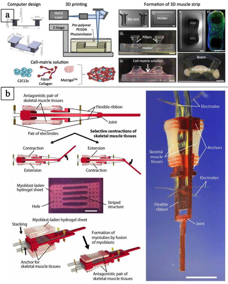
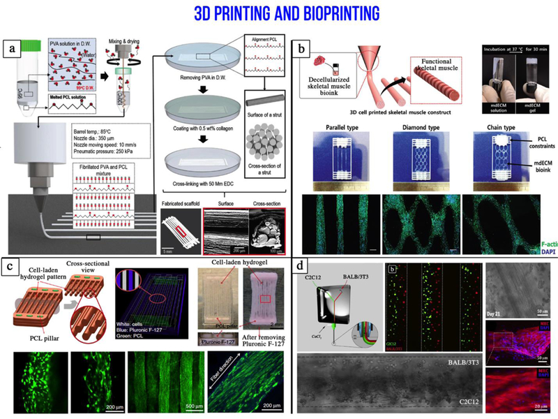
Skeletal muscle tissue engineering (SMTE) aims at repairing defective skeletal muscles. Until now, numerous developments are made in SMTE; however, it is still challenging to recapitulate the complexity of muscles with current methods of fabrication. Here, after a brief description of the anatomy of skeletal muscle and a short state-of-the-art on developments made in SMTE with "conventional methods," the use of 3D bioprinting as a new tool for SMTE is in focus. The current bioprinting methods are discussed, and an overview of the bioink formulations and properties used in 3D bioprinting is provided. Finally, different advances made in SMTE by 3D bioprinting are highlighted, and future needs and a short perspective are provided.

Keywords: 3D printing; bioinks; bioprinting; hydrogels; skeletal muscle; tissue engineering.


Figures

Figure 1.
Figure 1.
Skeletal muscle anatomy (Reproduced with permission from [1] ©2010 MedicalTerms.info)
Figure 2.
Figure 2.. Engineering skeletal muscle tissues by conventional and 3D bioprinting methods.
a) Engineering the myotendinous junction (MTJ) using 3D bioprinting. Myoblasts (C2C12) and fibroblasts (3T3) are precisely deposited to mimic the 3D organization of the native tissue (adapted with permission from [87] ©2015 IOP Publishing Ltd). b) Electrospun polymer gradient was used to engineer the MTJ: The mechanical stiffness increased along the gradient mimicking the interface between the tendon and skeletal muscle (adapted with permission from [135] ©2010 Elsevier Ltd). c) Engineering the neuromuscular junction (NMJ) using a microchip: The microchip allowed the co-culture of separate populations of motor neurons and human skeletal myoblasts connected through micro-channels (adapted with permission from [136] ©2018 Elsevier Ltd). d) Co-culture of muscle and embryonic bodies (EBs) of differentiated motor neurons on 3D printed hydrogel mold. The muscle-neuron tissue rings were then transferred to 3D printed bio-bot skeleton connecting two pillars (adapted with permission from [137] ©2017 Creative Commons Attribution 4.0 International License).
Figure 3.
Figure 3.. Three major bioprinting strategies:
(a) Micro-extrusion printers utilize direct mechanical or pneumatic dispensing systems to extrude continuous beads of cell-containing biomaterials. (b) Inkjet bioprinters use either a local pulsed joule heater to heat the print-head and produce air bubbles forcing droplets out of the nozzle, or a piezoelectric actuator to generate localized pressure via ultrasonic waves that can form droplets of bioink-cell hybrid. (c) Laser-assisted bioprinters (LAB) emit laser beams on an absorbing substrate that generate heat waves dispensing the cell-containing materials onto the substrate.
Figure 4.
Figure 4.
Percentage of materials used in skeletal muscle 3D printing over a period of 15 years from published articles. A) Cell-free and B) Cell-laden constructs. Adapted with permission from [99] ©2017 Future Medicine Ltd
Figure 5.
Figure 5.. Engineering biohybrid robots.
a-b) Assembling of biohybrid robots combining rapid prototyping techniques and living cells. a) 3D printed hydrogel “bio-bots” with an asymmetric physical design and powered by the actuation of an engineered mammalian skeletal muscle strip (adapted with permission from [101] ©2014 PNAS). b) Biohybrid robot powered by an antagonistic pair of skeletal muscle tissues (adapted with permission from Morimoto et al.[102] ©2018 American Association for the Advancement of Science).
Figure 6.
Figure 6.. Additive manufacturing in skeletal muscle tissue engineering.
a) Fabrication processes and optical/SEM images of the hybrid microfibrillated PCL/collagen scaffold used to mimic skeletal muscle hierarchical organization (adapted with permission from [107] ©2018 John Wiley and Sons). b) Three dimensional printing of an ink made from a decellularized (mdECM) porcine skeletal muscle to promote myoblast differentiation (adapted with permission from [109] ©2016 John Wiley and Sons). c) Integrated tissue-organ printer (ITOP) to fabricate human scale tissue constructs. Muscle precursors are encapsulated within hydrogel fibers that are supported by PCL pillars (adapted with permission from [108] ©2016 Springer Nature). d) Microfluidic printing head used to precisely deposit heterogeneous janus-like hydrogel fibers containing myoblasts and fibroblasts (adapted with permission from [93] ©2017 Elsevier).

References

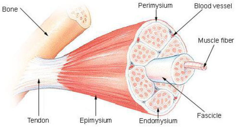
    1. M. T. E. O. GenericLook.com Skeletal muscle anatomy. http://medicalterms.info/anatomy/Skeletal-Muscles/.
    1. Bottinelli R, Reggiani C, Prog. Biophys. Mol. Biol. 2000, 73, 195. - PubMed
    1. Liu Y, Shen T, Randall WR, Schneider MF, J. Muscle Res. Cell Motil. 2005, 26, 13. - PubMed
    1. Sanger JW, Wang J, Fan Y, White J, Sanger JM, J. Biomed. Biotechnol. 2010. 2010, 1. - PMC - PubMed
    1. Ostrovidov S, Hosseini V, Ahadian S, Fujie T, Parthiban SP, Ramalingam M, Bae H, Kaji H, Khademhosseini A, Tissue Eng. Part B Rev. 2014, 20, 403. - PMC - PubMed

Publication types

MeSH terms

LinkOut - more resources