. 2019 Oct;71(10):1711-1722.
doi: 10.1002/art.40915. Epub 2019 Aug 27.

Induction of Inflammation and Fibrosis by Semaphorin 4A in Systemic Sclerosis

Affiliations

Induction of Inflammation and Fibrosis by Semaphorin 4A in Systemic Sclerosis

Tiago Carvalheiro et al. Arthritis Rheumatol. 2019 Oct.

Abstract

Objective: To analyze the potential role of semaphorin 4A (Sema4A) in inflammatory and fibrotic processes involved in the pathology of systemic sclerosis (SSc).

Methods: Sema4A levels in the plasma of healthy controls (n = 11) and SSc patients (n = 20) were determined by enzyme-linked immunosorbent assay (ELISA). The expression of Sema4A and its receptors in monocytes and CD4+ T cells from healthy controls and SSc patients (n = 6-7 per group) was determined by ELISA and flow cytometry. Th17 cytokine production by CD4+ T cells (n = 5-7) was analyzed by ELISA and flow cytometry. The production of inflammatory mediators and extracellular matrix (ECM) components by dermal fibroblast cells (n = 6) was analyzed by quantitative polymerase chain reaction, ELISA, Western blotting, confocal microscopy, and ECM deposition assay.

Results: Plasma levels of Sema4A, and Sema4A expression by circulating monocytes and CD4+ T cells, were significantly higher in SSc patients than in healthy controls (P < 0.05). Inflammatory mediators significantly up-regulated the secretion of Sema4A by monocytes and CD4+ T cells from SSc patients (P < 0.05 versus unstimulated SSc cells). Functional assays showed that Sema4A significantly enhanced the expression of Th17 cytokines induced by CD3/CD28 in total CD4+ T cells as well in different CD4+ T cell subsets (P < 0.05 versus unstimulated SSc cells). Finally, Sema4A induced a profibrotic phenotype in dermal fibroblasts from both healthy controls and SSc patients, which was abrogated by blocking or silencing the expression of Sema4A receptors.

Conclusion: Our findings indicate that Sema4A plays direct and dual roles in promoting inflammation and fibrosis, 2 main features of SSc, suggesting that Sema4A might be a novel therapeutic target in SSc.


Figures

Figure 1
Figure 1
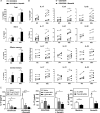
Semaphorin 4A (Sema4A) is elevated in plasma, monocytes, and CD4+ T cells from patients with systemic sclerosis (SSc) and is induced by inflammatory stimuli. A, Sema4A levels in plasma from healthy controls (HC; n = 11) and SSc patients (n = 20). Data are shown as box plots. Each box represents the 25th to 75th percentiles. Lines inside the boxes represent the median. Lines outside the boxes represent the 10th and 90th percentiles. B, Correlation between Sema4A plasma levels and the modified Rodnan skin thickness score (MRSS). C and D, Intracellular Sema4A expression in monocytes and CD4+ T cells from healthy controls (n = 6) and SSc patients (n = 6). Data are shown as the change in median fluorescence intensity (ΔMFI) (C) or the percentage of positive cells (D). E, Expression of Sema4A protein by monocytes from healthy controls and SSc patients (n = 6 per group). Cells were stimulated with poly(I‐C) or CXCL4 for 48 hours. F, Expression of Sema4A protein by CD4+ T cells from healthy controls and SSc patients (n = 6 per group). Cells were stimulated with CD3/CD28 Dynabeads for 5 days. G, Surface expression of plexin D1 and plexin B2 in CD4+ T cells from healthy controls and SSc patients. Circles represent individual subjects; horizontal lines and error bars show the mean ± SEM. H, Surface expression of plexin D1 and plexin B2 in CD4+ T cell subsets from healthy controls and SSc patients (n = 7 per group). I, Surface expression of plexin D1, plexin B2, and neuropilin 1 (NRP‐1) in total CD4+ T cells from healthy controls and SSc patients (n = 7 per group). Cells were stimulated with CD3/CD28 Dynabeads. In C–F, H, and I, bars show the mean ± SEM. * = P < 0.05; ** = P < 0.01; *** = P < 0.001 for the indicated comparisons. ## = P < 0.05 versus healthy control samples stimulated with poly(I‐C); $$ = P < 0.05 versus healthy control samples stimulated with CXCL4.
Figure 2
Figure 2
Semaphorin 4A (Sema4A)–induced Th17 cytokine production in CD4+ T cells. A and B, Interleukin‐17 (IL‐17) secretion (A) and intracellular levels of IL‐17, IL‐21, and IL‐22 (B) in total CD4+ T cells and different CD4+ T cell subsets from healthy controls (HCs) and patients with systemic sclerosis (SSc). Cells were activated with CD3/CD28 Dynabeads in the absence or presence of Sema4A for 5 days (for total CD4+ T cells), 7 days (for naive CD4+ T cells), or 2 days (for effector memory and central memory CD4+ T cells). C, Secretion of IL‐17 by total CD4+ T cells from SSc patients following 5 days of activation with CD3/CD28 Dynabeads in the absence or presence of Sema4A, which had previously been incubated for 1 hour with increasing concentrations of blocking anti–plexin D1 or anti–neuropilin 1 (anti–NRP‐1) antibodies or after plexin B2 silencing. In A and C, bars show the mean ± SEM of 5–7 independent experiments. In B, symbols represent individual subjects. * = P < 0.05; *** = P < 0.001 for the indicated comparisons. $ = P < 0.05 versus CD3/CD28‐activated cells from healthy controls; # = P < 0.05; ## = P < 0.01, versus CD3/CD28‐activated, Sema4A‐treated cells from healthy controls in A and versus medium or scrambled (Sc) small interfering RNA (siRNA) in C.
Figure 3
Figure 3
Semaphorin 4A (Sema4A) orchestrates fibroblast activation via interleukin‐17 (IL‐17) production by CD4+ T cells. A and B, Expression of mRNA for extracellular matrix components (A) and inflammatory mediators (B) in skin fibroblasts from healthy controls. Cells were incubated for 24 hours with conditioned medium of activated CD4+ T cells in the absence or presence of Sema4A, which had previously been incubated for 1 hour with an anti–IL‐17 antibody or its isotype control. C, Expression of IL‐6 and IL‐8 protein (ng/ml) by skin fibroblasts from healthy controls. Cells were incubated for 24 hours with conditioned medium of activated CD4+ T cells in the absence or presence of Sema4A, which had previously been incubated for 1 hour with an anti–IL‐17 antibody or its isotype control. Bars show the mean ± SEM of 7 independent experiments. * = P < 0.05 for the indicated comparisons. # = P < 0.05; ## = P < 0.01; ### = P < 0.001 versus medium.
Figure 4
Figure 4
Semaphorin 4A (Sema4A)–induced expression and deposition of extracellular matrix (ECM) components. A, Expression of mRNA for ECM components by skin fibroblasts from healthy controls (HCs) and patients with systemic sclerosis (SSc). Cells were stimulated with Sema4A (200 ng/ml) for 24, 48, or 72 hours. Bars show the mean ± SEM of 6 independent experiments. B, Representative immunoblots showing expression of type VI collagen (Col VI), type III collagen, vimentin, and α‐smooth muscle actin (α‐SMA) protein in skin fibroblasts from healthy controls and SSc patients. Cells were stimulated with Sema4A for 72 hours. C and D, Type I collagen, type IV collagen, and fibronectin 1 (FN1) production (C) and deposition (D) by skin fibroblasts from healthy controls and SSc patients. Cells were stimulated with Sema4A for 7 days. Images in C are representative of 4 independent experiments. Original magnification × 20. Symbols in D represent individual subjects. * = P < 0.05; ** = P < 0.01; *** = P < 0.001 for the indicated comparisons. # = P < 0.05 versus unstimulated healthy control fibroblasts.
Figure 5
Figure 5
Plexin D1 blocking and plexin B2 silencing abrogate semaphorin 4A (Sema4A)–induced expression of extracellular matrix (ECM) components. A and B, Expression of mRNA for ECM components (A) and representative immunoblots showing expression of type VI collagen (Col VI), type III collagen, vimentin, and α‐smooth muscle actin (α‐SMA) protein (B) by skin fibroblasts from patients with systemic sclerosis (SSc). Cells were stimulated with Sema4A for 72 hours after 1 hour of preincubation with blocking anti–plexin D1 antibody or its isotype control. C, Densitometric analysis of type III collagen, type VI collagen, vimentin, and α‐SMA protein expression. Data were normalized to histone H3 expression. D and E, Expression of mRNA for ECM components (D) and representative immunoblots showing expression of type VI collagen, type III collagen, vimentin, and α‐SMA protein (E) by skin fibroblasts from SSc patients. Cells were stimulated with Sema4A for 72 hours after plexin B2 silencing. F, Densitometric analysis of type III collagen, type VI collagen, vimentin, and α‐SMA protein expression. Data were normalized to histone H3 expression. In A, C, D, and F, bars show the mean ± SEM of 5–6 independent experiments. In B and E, results are representative of 5–6 independent experiments. * = P < 0.05; ** = P < 0.01; *** = P < 0.001 for the indicated comparisons. # = P < 0.05; ## = P < 0.01; ### = P < 0.001, versus isotype control or unstimulated scrambled (Sc) small interfering RNA (siRNA)–transfected cells.
Figure 6
Figure 6
Schematic overview of the inflammatory and fibrotic roles of semaphorin 4A (Sema4A) in the pathogenesis of systemic sclerosis (SSc). Sema4A is elevated in the plasma of SSc patients, due to increased production by monocytes and, to a lesser extent, CD4+ T cells. In CD4+ T cells, Sema4A enhances the production of Th17 cytokines induced by CD3/CD28, and secreted interleukin‐17 (IL‐17) induces the production of inflammatory mediators and chemokines in dermal fibroblasts. Sema4A also plays a direct role in fibrosis by inducing the production of extracellular matrix components and the expression of the myofibroblast marker α‐smooth muscle actin (α‐SMA) in dermal fibroblasts. NRP‐1 = neuropilin 1; Col I = type I collagen; COX‐2 = cyclooxygenase 2.

References

    1. Gabrielli A, Avvedimento EV, Krieg T. Scleroderma. N Engl J Med 2009;360:1989–2003. - PubMed
    1. Denton CP, Khanna D. Systemic sclerosis. Lancet 2017;390:1685–99. - PubMed
    1. Pattanaik D, Brown M, Postlethwaite BC, Postlethwaite AE. Pathogenesis of systemic sclerosis. Front Immunol 2015;6:272. - PMC - PubMed
    1. Ebmeier S, Horsley V. Origin of fibrosing cells in systemic sclerosis. Curr Opin Rheumatol 2015;27:555–62. - PMC - PubMed
    1. Van Bon L, Cossu M, Radstake TR. An update on an immune system that goes awry in systemic sclerosis. Curr Opin Rheumatol 2011;23:505–10. - PubMed

Publication types

MeSH terms