

Three-dimensional analysis of mandibular functional units in adult patients with unilateral posterior crossbite: A cone beam study with the use of mirroring and surface-to-surface matching techniques
- PMID: 31013132
- PMCID: PMC8117204
- DOI: 10.2319/081718-607.1
Three-dimensional analysis of mandibular functional units in adult patients with unilateral posterior crossbite: A cone beam study with the use of mirroring and surface-to-surface matching techniques
Abstract
Objectives: To use three-dimensional (3D) mirroring and surface-to-surface techniques to determine any differences in mandibular functional unit shape and morphology between the crossbite side and non-crossbite side in adult patients with posterior unilateral crossbite who had not received any corrective treatment for malocclusion.
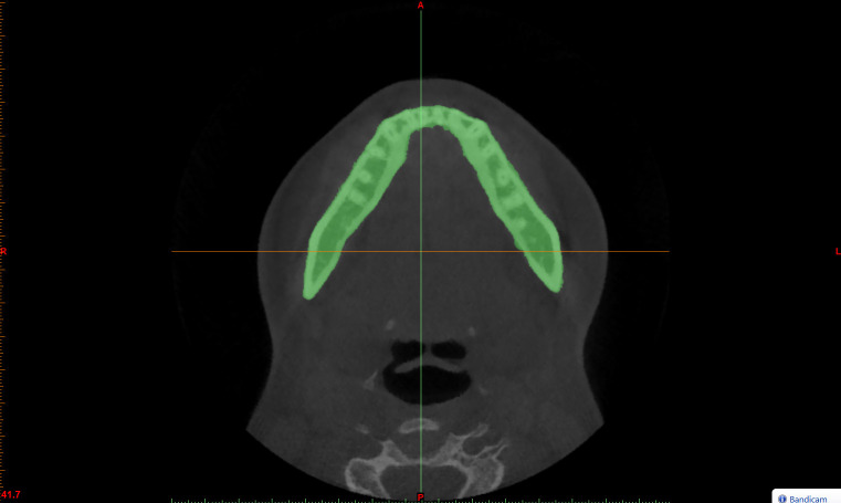
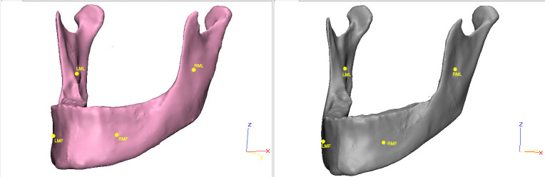
Materials and methods: Cone-beam computed tomography (CBCT) records from 24 consecutive adult white patients (mean age, 27.5 years; range 22.6-39.7 years; 14 women and 10 men) seeking treatment for maxillary transverse deficiency were assessed in this study. The control group comprised CBCT scans from age- and sex-matched patients. Segmentation masks were generated to obtain 3D surface mesh models of the mandibles and analyze the six skeletal functional units, which were further analyzed with reverse engineering software.
Results: Statistically significant differences in the mean surface distance when comparing the study sample and the control sample were found at the condylar process, mandibular ramus, angular process (P ≤ .0001), and alveolar process (P ≤ .01); no statistically significant differences were found for the coronoid process, the chin, and the mandibular body (P ≥ .5).
Conclusions: The condylar, angular, and alveolar processes plus the mandibular ramus appear to play a more dominant role than did the body, the coronoid, and the chin units in the asymmetry of the mandible in patients with unilateral crossbite.
Keywords: CBCT; Crossbite; Mandible; Surface-to-surface matching.
Figures


Similar articles
-
Evaluation of mandibular changes after rapid maxillary expansion: a CBCT study in youngsters with unilateral posterior crossbite using a surface-to-surface matching technique.Clin Oral Investig. 2021 Apr;25(4):1775-1785. doi: 10.1007/s00784-020-03480-5. Epub 2020 Aug 2. Clin Oral Investig. 2021. PMID: 32743674
-
Is there an asymmetry of the condylar and coronoid processes of the mandible in individuals with unilateral crossbite?Angle Orthod. 2019 May;89(3):464-469. doi: 10.2319/052518-398.1. Epub 2018 Dec 28. Angle Orthod. 2019. PMID: 30644758 Free PMC article.
-
Mandibular asymmetry in young adult patients with unilateral posterior crossbite: A controlled retrospective CBCT study.Int Orthod. 2021 Sep;19(3):433-444. doi: 10.1016/j.ortho.2021.05.003. Epub 2021 Jun 2. Int Orthod. 2021. PMID: 34088620
-
Mandibular asymmetry in unilateral and bilateral posterior crossbite patients using cone-beam computed tomography.Angle Orthod. 2011 Nov;81(6):966-74. doi: 10.2319/022011-122.1. Epub 2011 May 18. Angle Orthod. 2011. PMID: 21591969 Free PMC article.
-
Three-dimensional assessment of mandibular asymmetry in skeletal Class I and unilateral crossbite malocclusion in 3 different age groups.Am J Orthod Dentofacial Orthop. 2020 Aug;158(2):209-220. doi: 10.1016/j.ajodo.2019.08.010. Epub 2020 May 22. Am J Orthod Dentofacial Orthop. 2020. PMID: 32451206
Cited by
-
The Evolution of the Cephalometric Superimposition Techniques from the Beginning to the Digital Era: A Brief Descriptive Review.Int J Dent. 2021 Apr 23;2021:6677133. doi: 10.1155/2021/6677133. eCollection 2021. Int J Dent. 2021. PMID: 33981342 Free PMC article.
-
Evaluation of mandibular changes after rapid maxillary expansion: a CBCT study in youngsters with unilateral posterior crossbite using a surface-to-surface matching technique.Clin Oral Investig. 2021 Apr;25(4):1775-1785. doi: 10.1007/s00784-020-03480-5. Epub 2020 Aug 2. Clin Oral Investig. 2021. PMID: 32743674
-
Three-dimensional quantification of mandibular asymmetries in Caucasian adult patients with different sagittal and vertical skeletal patterns. A cone beam study using 3D segmentation and mirroring procedures.Head Face Med. 2023 Dec 14;19(1):54. doi: 10.1186/s13005-023-00400-2. Head Face Med. 2023. PMID: 38098053 Free PMC article.
-
External root resorption and rapid maxillary expansion in the short-term: a CBCT comparative study between tooth-borne and bone-borne appliances, using 3D imaging digital technology.BMC Oral Health. 2023 Aug 12;23(1):558. doi: 10.1186/s12903-023-03280-9. BMC Oral Health. 2023. PMID: 37573295 Free PMC article.
-
One Step before 3D Printing-Evaluation of Imaging Software Accuracy for 3-Dimensional Analysis of the Mandible: A Comparative Study Using a Surface-to-Surface Matching Technique.Materials (Basel). 2020 Jun 21;13(12):2798. doi: 10.3390/ma13122798. Materials (Basel). 2020. PMID: 32575875 Free PMC article.
References
-
- Kutin G, Hawes RR. Posterior cross-bites in the deciduous and mixed dentitions. Am J Orthod. 1969;56:491–504. - PubMed
-
- Zhu JF, Crevoisier R, King DL, Henry R, Mills CM. Posterior crossbites in children. Compend Contin Educ Dent. 1996;17:1051–1054. 1056, 1058. - PubMed
-
- Pinto AS, Buschang PH, Throckmorton GS, P Chen. Morphological and positional asymmetries of young children with functional unilateral posterior crossbite. Am J Orthod Dentofacial Orthop. 2001;120:513–520. - PubMed
-
- Tsanidis N, Antonarakis GS, Kiliaridis S. Functional changes after early treatment of unilateral posterior cross-bite associated with mandibular shift: a systematic review. J Oral Rehabil. 2016;43:59–68. - PubMed
MeSH terms
LinkOut - more resources
Full Text Sources
