. 2019 Apr 22;20(8):1956.
doi: 10.3390/ijms20081956.

Disruption of Structural Disulfides of Coagulation FXIII-B Subunit; Functional Implications for a Rare Bleeding Disorder

Affiliations

Disruption of Structural Disulfides of Coagulation FXIII-B Subunit; Functional Implications for a Rare Bleeding Disorder

Sneha Singh et al. Int J Mol Sci. .

Abstract

Congenital FXIII deficiency is a rare bleeding disorder in which mutations are detected in F13A1 and F13B genes that express the two subunits of coagulation FXIII, the catalytic FXIII-A, and protective FXIII-B. Mutations in FXIII-B subunit are considerably rarer compared to FXIII-A. Three mutations in the F13B gene have been reported on its structural disulfide bonds. In the present study, we investigate the structural and functional importance of all 20 structural disulfide bonds in FXIII-B subunit. All disulfide bonds were ablated by individually mutating one of its contributory cysteine's, and these variants were transiently expressed in HEK293t cell lines. The expression products were studied for stability, secretion, the effect on oligomeric state, and on FXIII-A activation. The structural flexibility of these disulfide bonds was studied using classical MD simulation performed on a FXIII-B subunit monomer model. All 20 FXIII-B were found to be important for the secretion and stability of the protein since ablation of any of these led to a secretion deficit. However, the degree of effect that the disruption of disulfide bond had on the protein differed between individual disulfide bonds reflecting a functional hierarchy/diversity within these disulfide bonds.

Keywords: FXIII deficiency; FXIII-B; coagulation Factor XIII; disulfide bonds.


Conflict of interest statement

The authors declare no conflict of interest.

Figures

Figure 1
Figure 1
Transient expression of FXIII-B Cysteine mutants. Panel A. A conventional western blot of 10uL each of the wild type FXIII-B and 20 rFXIII-B cysteine mutants retained from the culture medium of transfected cells, probed against mouse to human FXIII-B antibody. Lanes: 1: Un-transfected control; 2: C76A; 3: Wild-Type; 4: C364A; 5: C396A; 6: C425A; 7: C454A; 8: C524A; 9: C553A; 10: C582A; 11: C616A; 12: C486A; 13: C378A; 14: C59A; 15: C91A; 16: C118A; 17: C153A; 18: C180A; 19: C213A; 20:C267A; 21:C274A; 22: C302A; and 23: rFXIII-B (Zedira, 75ng, positive control) White arrows represent the wild-type FXIII-B. Panel B. A comparative bar-plot representation of the antigenic levels of FXIII-B cysteine mutants versus the FXIII-B wild type evaluated on a quantitative sandwich ELISA based platform, detecting FXIII-B in 100µL of sample retained from transiently transfected cells. Green and red bars represent the intracellular and the extracellular fractions, respectively. A “*” symbol represents significance (p-value < 0.05).
Figure 2
Figure 2
Effect of FXIII-B cysteine mutations on intracellular trafficking of FXIII-B protein. Panel A. Confocal microscopy tracking the subcellular localization of FXIII-B cysteine mutant proteins, via cell-specific markers; green (αFXIII-B), red (α-PDI (for endoplasmic reticulum) and α-TGN-46 (for trans-Golgi network)) with secondary antibodies conjugated with Alexa-488 and Alexa-555 respectively. Bars represent 10µm scale. Panel B. Bar-plot representation of Pearson’s coefficient calculated as a measurement of the intensity of pixels, defining co-localization of FXIII-B within either endoplasmic reticulum (PDI), or trans-GGolgi network (TGN-46). A “*” symbol represents significance (p-value < 0.05). Panel C. Bar plot representation of antigenic evaluation (ELISA) of select FXIII-B cysteine mutants which showed higher intracellular retention (on confocal immunostaining) when compared to wild-type. A “*” symbol represents significance (p-value < 0.05).
Figure 3
Figure 3
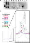
Altered complexation/Oligomerization of FXIII-B Cys mutants. Panel A. Western blot of rFXIII-B cysteine mutants after separation by native PAGE which were found to be secreted successfully (as detected by ELISA). Lanes: 1: Wild-Type; 2: C153A; 3: C118A; 4: C180A; 5: C267A; 6: C274A; 7: C396A; 8: C425A; 9: C454A; 10: C616A; and 11: rFXIII-B (Zedira, 75ng); 12: C302A. Vertical white arrows represent the Wild-type and rFXIII-B (Zedira GmbH, Darmstadt, Germany), whereas horizontal white arrows here indicate the diverse mobility of protein bands corresponding to FXIII-B (since the western blot has been probed with the mouse to human FXIII-B antibodies) Panel B. Gel-filtration chromatography of purified FXIII-B cysteine mutants. Color codes represent respective mutants indicated as inset in the figure. The x-axis denotes retention volume [ml], and the y-axis represents the amount of protein in mAU (UV-280nm).
Figure 4
Figure 4
The FXIII-B subunit monomer model. Panel A shows the FXIII-B subunit monomer post-equilibrated model structure in ribbon format. The backbone is colored based on secondary structure. The disulfide-bonded cysteines are represented as grey colored stick forms. The individual sushi domains are numbered S1 to S10; N to C terminal. Panel B is the electrostatic surface representation of the FXIII-B subunit monomer model. The left side of Panel B shows the PBS styled depiction of surface electrostatics while the right side shows the PME styled depiction of surface electrostatic. Calculation and depiction for both forms of electrostatics were performed with macros embedded in YASARA. Red color indicates negative potential while blue represents positive potential.
Figure 5
Figure 5
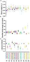
Evaluation of flexibility and stability of the disulfide bonds in the FXIII-B subunit monomer model. Panel A shows the variation in disulfide bond lengths during the simulation post equilibration of the FXIII-B subunit monomer model. Panel B shows the variation in disulfide strain energies of all disulfide bonds of the FXIII-B subunit monomer model during the MD simulation post-equilibration of the structure. Panel C shows the variability in free energy change when individual disulfide-bonded cysteines (the cysteines that have been mutated and expressed in this study) within the FXIII-B subunit monomer model are mutated to alanine. The evaluation was performed on several simulation trajectory structures (spaced at an interval of 20 ns) within the post-equilibrated simulation.
Figure 6
Figure 6
Effect of cysteine mutants on the rate of FXIII-Aa generation. This figure represents the comparative bar-plot representation of FXIII-Aa generation assay. The maximum rate of FXIII-Aa generation (µ) (in ∆RFU/min) upon spiking of FXIII-deficient plasma with 7 µg/mL rFXIII-A2 is represented in the x-axis, in the absence (grey bar), or presence (color-coded bars) of rFXIII-B2 subunits or FXIII-B cysteine mutants (10µg/mL each).

Similar articles

Cited by

References

    1. Biswas A., Ivaskevicius V., Thomas A., Oldenburg J. Coagulation factor XIII deficiency. Diagnosis, prevalence and management of inherited and acquired forms. Hamostaseologie. 2014;34:160–166. - PubMed
    1. Gupta S., Biswas A., Akhter M.S., Krettler C., Reinhart C., Dodt J., Reuter A., Philippou H., Ivaskevicius V., Oldenburg J. Revisiting the mechanism of coagulation factor XIII activation and regulation from a structure/functional perspective. Sci. Rep. 2016;6:30105. - PMC - PubMed
    1. Duckert F., Jung E., Shmerling D.H. A hitherto undescribed congenital haemorrhagic diathesis probably due to fibrin stabilizing factor deficiency. Thromb. et Diath. Haemorrh. 1960;5:179–186. doi: 10.1055/s-0038-1654918. - DOI - PubMed
    1. Seitz R., Duckert F., Lopaciuk S., Muszbek L., Rodeghiero F., Seligsohn U. ETRO Working Party on Factor XIII questionnaire on congenital factor XIII deficiency in Europe: Status and perspectives. Study Group. Semin. Thromb. and Hemost. 1996;22:415–418. doi: 10.1055/s-2007-999040. - DOI - PubMed
    1. Biswas A., Ivaskevicius V., Seitz R., Thomas A., Oldenburg J. An update of the mutation profile of Factor 13 A and B genes. Blood Rev. 2011;25:193–204. doi: 10.1016/j.blre.2011.03.001. - DOI - PubMed

LinkOut - more resources