Review
. 2019 Apr 22;24(8):1578.
doi: 10.3390/molecules24081578.

Molecular Recognition of the Hybrid-Type G-Quadruplexes in Human Telomeres

Affiliations
Review

Molecular Recognition of the Hybrid-Type G-Quadruplexes in Human Telomeres

Guanhui Wu et al. Molecules. .

Abstract

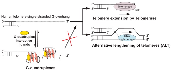
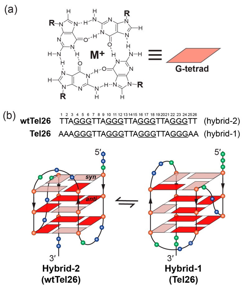
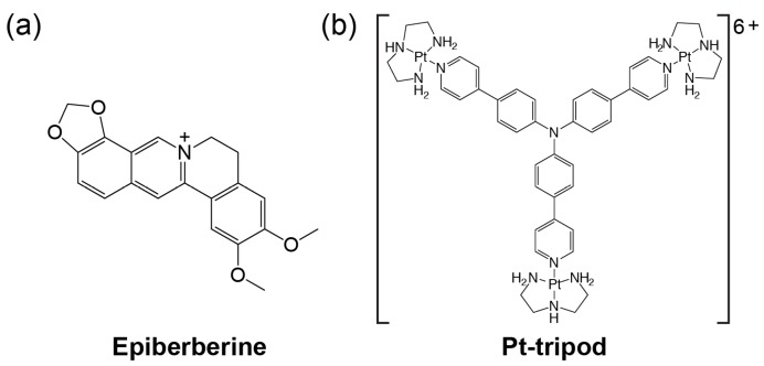
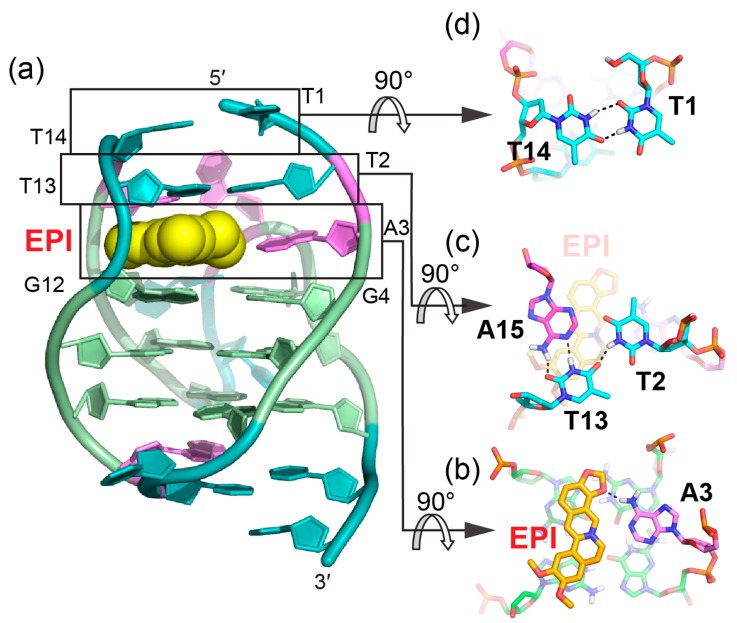
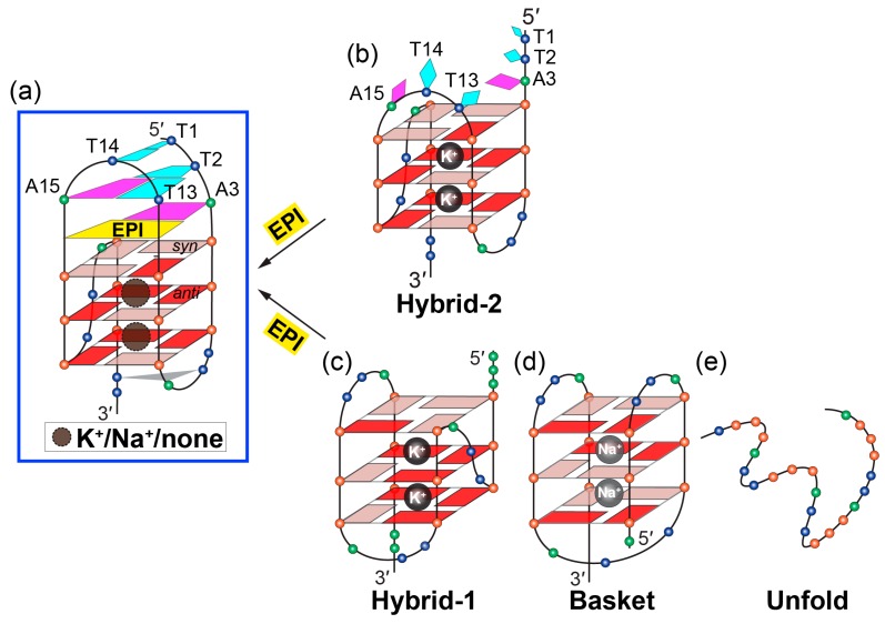
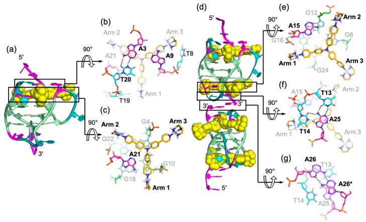
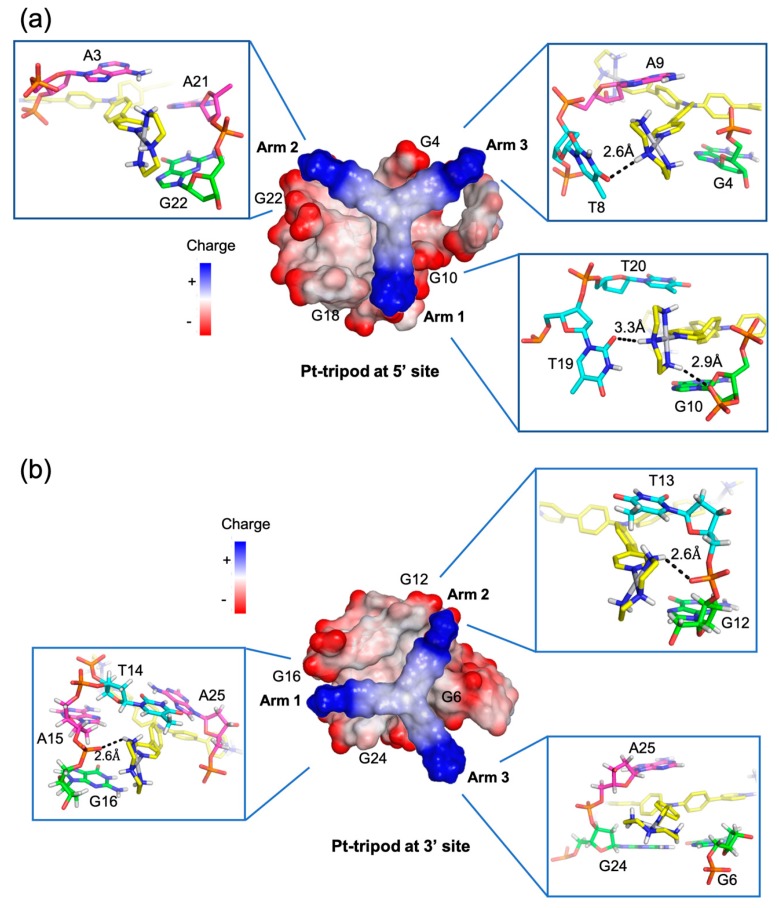
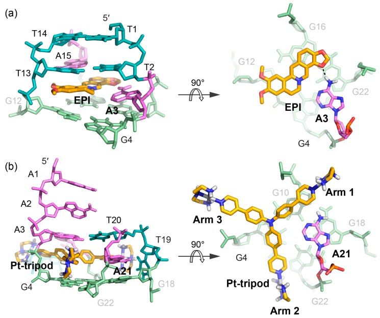
G-quadruplex (G4) DNA secondary structures formed in human telomeres have been shown to inhibit cancer-specific telomerase and alternative lengthening of telomere (ALT) pathways. Thus, human telomeric G-quadruplexes are considered attractive targets for anticancer drugs. Human telomeric G-quadruplexes are structurally polymorphic and predominantly form two hybrid-type G-quadruplexes, namely hybrid-1 and hybrid-2, under physiologically relevant solution conditions. To date, only a handful solution structures are available for drug complexes of human telomeric G-quadruplexes. In this review, we will describe two recent solution structural studies from our labs. We use NMR spectroscopy to elucidate the solution structure of a 1:1 complex between a small molecule epiberberine and the hybrid-2 telomeric G-quadruplex, and the structures of 1:1 and 4:2 complexes between a small molecule Pt-tripod and the hybrid-1 telomeric G-quadruplex. Structural information of small molecule complexes can provide important information for understanding small molecule recognition of human telomeric G-quadruplexes and for structure-based rational drug design targeting human telomeric G-quadruplexes.

Keywords: G-quadruplex; G4; anticancer drug; epi-berberine; human telomeres; hybrid-1; hybrid-2; molecular recognition; platinum-tripod; rational drug design; solution structure.


Conflict of interest statement

The authors declare no conflict of interest.

Figures

Figure 1
Figure 1
Biological implications of targeting telomeric G-quadruplexes using G-quadruplex-interactive ligands. In human tumors, the free single-stranded telomere G-overhang can be extended by telomerase, which reverse transcribes the template region of its RNA subunit (hTR), or by alternative lengthening of telomeres (ALT), which copies telomeric template DNA via homologous recombination. G-quadruplex-interactive ligands can promote the formation of and stabilize the telomeric G-quadruplex structures, thus inhibiting telomere extension processes which are required for the indefinite growth of human tumors.
Figure 2
Figure 2
Schematic illustration of G-tetrad and the folding pattern of hybrid-2 and hybrid-1 telomeric G-quadruplexes. (a) Schematic representation of G-tetrad. Monovalent cations (M+), such as K+ or Na+, are required for the G-tetrad formation. (b) Human telomeric sequences predominantly fold into two hybrid-type G-quadruplexes with an equilibrium between hybrid-1 (PDB ID: 2HY9) and hybrid-2 (PDB ID: 2JPZ) forms. The human telomeric sequences used to determine hybrid-2 (wtTel26) and hybrid-1 (Tel26) structures are shown on the top. Strand polarities are indicated as the black arrows. Glycosidic conformations of guanine nucleotides are marked as follows: syn-pink and anti-red. Different nucleotides are represented as follows: Thymine, blue sphere; guanine, red sphere; and adenine, green sphere.
Figure 3
Figure 3
The chemical structures of epiberberine (a) and Pt-tripod (b).
Figure 4
Figure 4
The specific recognition of human hybrid-2 telomeric G-quadruplex by EPI (PDB ID: 6CCW) (a) Cartoon representation of 1:1 EPI/hybrid-2 complex. Different nucleotides are marked as follows: Thymine, cyan; adenine, pink; and guanine, green. The EPI molecule is shown as the yellow spheres. (bd) Top view of the EPI:A3 quasi-triad plane (b), T2:T13:A15 triad plane (c), and T1:T14 pair (d). Potential hydrogen bonds are shown in black dashed lines.
Figure 5
Figure 5
Conversion of human telomeric G-quadruplex structures to the hybrid-2 form induced by EPI. (ab) EPI binding induces extensive rearrangement of previously disordered 5′-flanking and lateral loop segments (b) to form a well-defined four-layer binding pocket (a) specific to hybrid-2 telomeric G-quadruplex. (ce) Other human telomeric G-quadruplex forms, including hybrid-1 (c) and basket (d), or the free telomeric sequence in the absence of salt (e) can be converted to the hybrid-2 form as the addition of EPI molecules. The specific recognition of human hybrid-2 telomeric G-quadruplex by EPI (PDB ID: 6CCW).
Figure 6
Figure 6
Solution structure of 1:1 and 4:2 Pt-tripod/hybrid-1 G-quadruplex complexes (PDB ID: 5Z80 and 5Z8F). (a) Cartoon representation of 1:1 Pt-tripod/hybrid-1 complex. Different nucleotides are marked as follows: Thymine, cyan; adenine, pink; and guanine, green. The Pt-tripod molecule is shown as the yellow spheres. (bc) Top views of the A3:A9:T20 triad plane (b) and Pt-tripod:A21 plane at 5′ site (c). (d) Cartoon representation of 4:2 Pt-tripod/hybrid-1 complex. (eg) Structural details of the 3′ site binding pocket. Bottom views of the Pt-tripod:A15 plane (e), T13:T14:A25 triad plane (f), and the A26:A26* (A26 from each hybrid-1) base pair (g). Potential hydrogen bonds are shown in black dashed lines.
Figure 7
Figure 7
Arm-groove interactions of Pt-tripod at 5′ and 3′ sites in the 4:2 Pt-tripod/hybrid-1 complex. (a) Top view of Pt-tripod at the 5′ site of hybrid-1 without capping structure (middle). The binding pocket surface is color coded according to the charge. The interaction details between each arm and groove are shown in enlarged view (corners). (b) The same information is shown for Pt-tripod at the 3′ site. Potential hydrogen bonds are shown in black dashed lines.
Figure 8
Figure 8
Comparison of the 5′-binding modes between EPI and Pt-tripod with human telomeric hybrid-type G-quadruplexes (a) Upon binding, the 5′-flanking A3 is recruited by EPI and forms a quasi-triad plane (right). The multi-layer drug-binding pocket for EPI is formed by the 5′-end flanking residues and TTA lateral loop. (left). Potential hydrogen bond is shown as the black dashed line. (b) Upon binding, A21 in the second lateral loop is recruited by Pt-tripod and forms a quasi-triad plane (right), stacking on top of the 5′-external G-tetrad and locking the position of Pt-tripod. Each arm of Pt-tripod stretches into different grooves. The Pt-tripod:A21 plane is capped by the A3:A9:T20 triad (left).

Similar articles

Cited by

References

    1. Verdun R.E., Karlseder J. Replication and protection of telomeres. Nature. 2007;447:924–931. doi: 10.1038/nature05976. - DOI - PubMed
    1. O’Sullivan R.J., Karlseder J. Telomeres: Protecting chromosomes against genome instability. Nat. Rev. Mol. Cell Biol. 2010;11:171–181. doi: 10.1038/nrm2848. - DOI - PMC - PubMed
    1. Hackett J.A., Feldser D.M., Greider C.W. Telomere dysfunction increases mutation rate and genomic instability. Cell. 2001;106:275–286. doi: 10.1016/S0092-8674(01)00457-3. - DOI - PubMed
    1. Vaziri H., Dragowska W., Allsopp R.C., Thomas T.E., Harley C.B., Lansdorp P.M. Evidence for a mitotic clock in human hematopoietic stem cells: Loss of telomeric DNA with age. Proc. Nat. Acad. Sci. USA. 1994;91:9857–9860. doi: 10.1073/pnas.91.21.9857. - DOI - PMC - PubMed
    1. Maciejowski J., de Lange T. Telomeres in cancer: Tumour suppression and genome instability. Nat. Rev. Mol. Cell Biol. 2017;18:175–186. doi: 10.1038/nrm.2016.171. - DOI - PMC - PubMed

MeSH terms

LinkOut - more resources