. 2019 Apr 22;20(8):1967.
doi: 10.3390/ijms20081967.

Involvement of p38 Activation and Mitochondria in Death of Human Leukemia Cells Induced by an Agonistic Human Monoclonal Antibody Fab Specific to TRAIL Receptor 1

Affiliations

Involvement of p38 Activation and Mitochondria in Death of Human Leukemia Cells Induced by an Agonistic Human Monoclonal Antibody Fab Specific to TRAIL Receptor 1

You-Ri Lee et al. Int J Mol Sci. .

Abstract

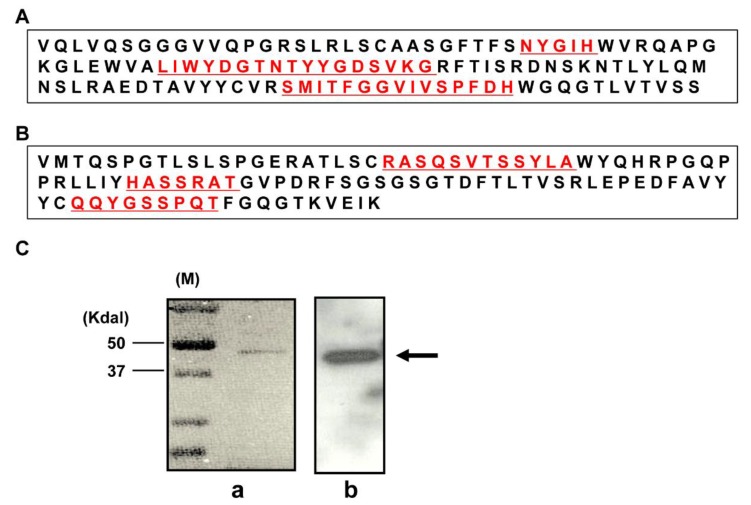
The tumor necrosis factor-related apoptosis-inducing ligand (TRAIL) induces cancer cell death with minimal damage to normal cells; however, some cancer cells are resistant to TRAIL. TRAIL resistance may be overcome by agonistic antibodies to TRAIL receptors. In this study, we report the toxic effects of a novel recombinant agonistic human anti-TRAIL receptor 1 (DR4) monoclonal antibody Fab fragment, DR4-4, on various TRAIL-resistant and -sensitive cancer cell lines. The mechanisms of DR4-4 Fab-induced cell death in a human T cell leukemia cell line (Jurkat) were investigated using cell viability testing, immunoblotting, immunoassays, flow cytometry, and morphological observation. DR4-4 Fab-induced caspase-independent necrosis was observed to occur in Jurkat cells in association with p38 mitogen-activated protein kinase activation, cellular FLICE (FADD-like IL-1β-converting enzyme)-inhibitory protein degradation, decreased mitochondrial membrane potential, and increased mitochondrial reactive oxygen species production. Increased cytotoxic effects of DR4-4 Fab were observed in combination with TRAIL or γ-irradiation. Our results indicate that the novel DR4-4 Fab might overcome TRAIL-resistance and induce death in leukemia cells via cellular mechanisms different from those activated by TRAIL. DR4-4 Fab may have application as a potential therapeutic antibody fragment in single or combination therapy for cancer.

Keywords: DR4; Fab; TRAIL; TRAIL receptor 1; TRAIL-resistance; agonistic human antibody; cancer cells; mitochondria; p38.


Conflict of interest statement

The authors declare no conflict of interest.

Figures

Figure 1
Figure 1
Amino acid sequences of heavy (VH) and light (VL) chains and visualization of the purified DR4-4 Fab. The amino acid sequences of the VH (A) and VL (B) regions of DR4-4 Fab are available from European Molecular Biology Laboratory/GenBank under accession numbers JN030159 (VH) and JN030158 (VL). The purified DR4-4 Fab (1 µg/mL for immunoblotting and 10 µg/mL for Coomassie blue staining) was visualized by sodium dodecyl sulfate-polyacrylamide gel electrophoresis (SDS-PAGE) and immunoblotting with anti–human IgG (Fab specific) monoclonal antibody (Ca) and Coomassie blue staining (Cb). DR4-4 Fab is presented by an arrow at approximately 45 kDa.
Figure 2
Figure 2
Specific binding of DR4-4 Fab to DR4 antigen. Direct-binding (A) and competitive (B) enzyme-linked immunosorbent assay (ELISA) for specific binding of DR4-4 Fab to DR4. Recombinant DR4 and DR5 were coated onto the wells of ELISA plates at 5 µg/mL, followed by incubation with DR4-4 Fab (A) or DR4-4 Fab preincubated with competitor, DR4 or DR5 (B) (data presented as mean ± standard deviation). (C) Binding affinity of recombinant human DR4 antigen for DR4-4 (1 µM) immobilized on a nitrile triacetic acid chip as measured by Biacore surface plasmon resonance. (D) Fluorescence-activated cell sorting analysis of the cellular binding of DR4-4 Fab. Cells (5 × 10⁵) were incubated with fluorescein isothiocyanate (FITC)-labeled DR4-4 Fab for 30 min at 4 °C without (a) or with (b) pretreatment with unlabeled DR4-4 Fab. Dot plot presents the profile of forward scatter (FSC)/side scatter (SSC) of control cells. P is a gate of cells which were used for the analysis. (C,D) are representative results among triplicate experiments.
Figure 3
Figure 3
Specific cytotoxic effects of DR4-4 Fab on cancer cells. Cells (2.5 × 10⁴) were treated with the indicated concentrations of TRAIL (A) or DR4-4 Fab (B) for 48 h. (C) Viability (as assessed by MTT assay) of normal human fibroblasts (1 × 10⁵) treated with TRAIL or DR4-4 Fab for 48 h. (D) Cell morphology of normal human fibroblasts treated with TRAIL or DR4-4 Fab for 48 h was evaluated by phase contrast microscopy at 100×. Data presented as mean ± standard deviation.
Figure 4
Figure 4
Caspase-independent and mitochondrial-induced cytotoxicity of DR4-4 Fab. (A) Jurkat cells (2.5 × 10⁴) were treated with TRAIL (125 ng/mL) or DR4-4 Fab (100 µg/mL) for the indicated time and cell viability was measured by MTT assay. (B) Cells (1 × 10⁶) were treated with TRAIL (125 ng/mL) or DR4-4 Fab (100 µg/mL) for the indicated time with or without a 3 h pretreatment with z-VAD (20 µM). Caspase-3, cleaved caspase-3, poly(ADP-ribose) polymerase (PARP), and cleaved PARP were detected by immunoblotting with specific antibodies. β-actin was used as a control to quantify the relative amount of sample loaded in each well. (C) MTT assay after treatment of cells with DR4-4 Fab (30 min pretreatment with or without z-VAD (20 µM), Nec-1 (50 µM), and N-acetyl-l-cysteine (NAC; 5 mM)) or TRAIL (30 min pretreatment with or without z-VAD (20 µM)). (D) Cells were treated with DR4-4 Fab (100 µg/mL) for 6 h in the presence or absence of NAC (5 mM), stained with annexin V-FITC/PI, and analyzed by flow cytometry (1 × 10⁶ cells were counted). (E) Mitochondrial reactive oxygen species generation was measured by flow cytometry after treatment with DR4-4 Fab (100 µg/mL) for the indicated time (0–48 h) and 5 µM MitoSOX staining. (F) Cells were treated with 100 µg/mL DR4-4 Fab (0, 16, 24, 32, and 48 h) and stained with JC-1 to analyze mitochondrial membrane potential (MMP). The ratio of red to green forms of JC-1 was measured by flow cytometry. (G) Cells were treated with 100 µg/mL DR4-4 Fab for 48 h with or without wortmannin (WM) and 1,2-bis(o-aminophenoxy)ethane-N,N,N′,N′-tetraacetic acid (BAPTA; 10 µM). After staining with JC-1, cells were analyzed by flow cytometry for MMP. Data presented as mean ± standard deviation (* p < 0.05, ** p < 0.005, versus control).
Figure 5
Figure 5
Morphological observation of cells treated with DR4-4 Fab. (A) Representative transmission electron microscopy images taken after THP-1 cell treatment with 50 µg/mL DR4-4 Fab for 0 h (a), 24 h (b), or 48 h (c,d). Scale bar, 2.5 µm. (B) Representative images showing morphological changes in the mitochondrial network of HeLa cells treated with 50 µg/mL DR4-4 Fab for 24 h observed by confocal microscopy (100×) after MitoTracker Red FM staining of mitochondria. Scale bar, 5 µm. The parts of the images were enlarged and presented in yellow squares.
Figure 6
Figure 6
Immunoblot analysis of signaling molecules induced by DR4-4 Fab. Immunoblotting performed after the indicated length of treatment of Jurkat cells (1 × 10⁶) with TRAIL (125 ng/mL) or DR4-4 Fab (100 µg/mL). (A) P-p38 and p38 were detected with specific antibodies. (B) P-JNK and IκBα were detected after the treatment of TRAIL or DR4-4 Fab. P-ERK and ERK were analyzed only after the treatment with DR4-4 Fab. (C) C-FLIP, tBID, Bcl-xL, Bcl-2, Mcl-1, and Bax were analyzed with specific antibodies after the treatment of TRAIL or DR4-4 Fab. α-tubulin and β-actin were used to quantify the relative amount of sample loaded into each well. Bax, Bcl-2-associated X protein; Bcl-2, B-cell lymphoma 2; Bcl-xL, B-cell lymphoma extra-large; c-FLIP, cellular FADD-like IL-1β-converting enzyme–inhibitory protein; ERK, extracellular signal-regulated kinase; IκBα, nuclear factor κB inhibitor α; JNK, c-Jun N-terminal kinase; Mcl-1, myeloid cell leukemia 1; p-, phosphorylated protein; t-Bid, truncated cytosolic BH3 interacting-domain death agonist.
Figure 7
Figure 7
Enhanced rates of cell death following cotreatment with DR4-4 Fab and TRAIL or γ-irradiation. (A) Cytotoxicity of cotreatment with DR4-4 Fab and TRAIL as measured by MTT assay. Cell viability for single treatment with TRAIL is also presented with concentrations indicated (upmost line). (B) Cells pre-irradiated with γ-radiation (absorbed dose = 2 Gy) were treated with DR4-4 Fab (100 µg/mL) for 24 h. Cell death was measured by MTT assay. Data presented as mean ± standard deviation (* p < 0.05, ** p < 0.005, versus control).

Similar articles

References

    1. Azijli K., Yuvaraj S., van Roosmalen I., Flach K., Giovannetti E., Peters G., de Jong S., Kruyt F. MAPK p38 and JNK have opposing activities on TRAIL-induced apoptosis activation in NSCLC H460 cells that involves RIP1 and caspase-8 and is mediated by Mcl-1. Apoptosis. 2013;18:851–860. doi: 10.1007/s10495-013-0829-3. - DOI - PubMed
    1. Bellail A., Qi L., Mulligan P., Chhabra V., Hao C. TRAIL Agonists on Clinical Trials for Cancer Therapy: The Promises and the Challenges. Rev. Recent Clin. Trials. 2009;4:34–41. doi: 10.2174/157488709787047530. - DOI - PubMed
    1. Jang Y.-J., Park K., Chung H., Kim H. Analysis of the phenotypes of Jurkat clones with different TRAIL-sensitivities. Cancer Lett. 2003;194:107–117. doi: 10.1016/S0304-3835(02)00680-8. - DOI - PubMed
    1. Griffith T.S., Rauch C.T., Smolak P.J., Waugh J.Y., Boiani N., Lynch D.H., Smith C.A., Goodwin R.G., Kubin M.Z. Functional analysis of TRAIL receptors using monoclonal antibodies. J. Immunol. 1999;162:2597–2605. - PubMed
    1. Rowinsky E.K. Targeted induction of apoptosis in cancer management: The emerging role of tumor necrosis factor-related apoptosis-inducing ligand receptor activating agents. J. Clin. Oncol. 2005;23:9394–9407. doi: 10.1200/JCO.2005.02.2889. - DOI - PubMed

MeSH terms

Substances

LinkOut - more resources