. 2019 Apr 12;10(4):300.
doi: 10.3390/genes10040300.

Molecular Characterization of a Dirofilaria immitis Cysteine Protease Inhibitor (Cystatin) and Its Possible Role in Filarial Immune Evasion

Affiliations

Molecular Characterization of a Dirofilaria immitis Cysteine Protease Inhibitor (Cystatin) and Its Possible Role in Filarial Immune Evasion

Xiaowei Dong et al. Genes (Basel). .

Abstract

Infection with canine heartworm (Dirofilaria immitis), spread via mosquito vectors, causes coughing, asthma, pneumonia, and bronchitis in humans and other animals. The disease is especially severe and often fatal in dogs and represents a serious threat to public health worldwide. Cysteine protease inhibitors (CPIs), also known as cystatins, are major immunomodulators of the host immune response during nematode infections. Herein, we cloned and expressed the cystatin Di-CPI from D. immitis. Sequence analysis revealed two specific cystatin-like domains, a Q-x-V-x-G motif, and a SND motif. Phylogenetic analysis indicates that Di-CPI is a member of the second subgroup of nematode type II cystatins. Probing of D. immitis total proteins with anti-rDi-CPI polyclonal antibody revealed a weak signal, and immunofluorescence-based histochemical analysis showed that native Di-CPI is mainly localized in the cuticle of male and female worms and the gut of male worms. Treatment of canine peripheral blood mononuclear cells (PMBCs) with recombinant Di-CPI induced a Th2-type immune response characterized by high expression of the anti-inflammatory factor interleukin-10. Proliferation assays showed that Di-CPI inhibits the proliferation of canine PMBCs by 15%. Together, the results indicate that Di-CPI might be related to cellular hyporesponsiveness in dirofilariasis and may help D. immitis to evade the host immune system.

Keywords: Dirofilaria immitis; cystatin; immune evasion; immunohistochemistry; peripheral blood mononuclear cells.


Conflict of interest statement

The authors declare that the research was conducted in the absence of any commercial or financial relationships that could be construed as a potential conflict of interest.

Figures

Figure 1
Figure 1
Sequence alignment of Dirofilaria immitis cysteine protease inhibitor (Di-CPI). Alignment of the deduced Di-CPI amino acid sequence with those of homologous proteins from nematodes (Onchocerca volvulus, AAA29423.1; Brugia malayi, AAD51086.1; Litomosoides sigmodontis, AAF35896.1; Loa loa, XP_003147913; Acanthocheilonema viteae, AAA87228.1) and mammals (Mus musculus, NP_034106.2; Human, AAH13083.1; Canine, XP_003639869.1) was performed using Clustal X software version 2 and shaded using BOXshade version 3.21. Predicted secondary structural elements, including 
strands and helices, are shown above the alignment as arrows and loops,
respectively. The Q-x-V-x-G motif and SND motif are enclosed in red boxes. A conserved glycine residue in the N-terminal region and the PW hairpin loop in the C-terminal region are enclosed in the blue boxes. N-terminal signal peptides are enclosed in a red box.
Figure 2
Figure 2
Phylogenetic relationships between Di-CPI and homologous cystatins/CPIs. The tree was constructed from a multiple sequence alignment performed using Clustal W2 and plotted using MEGA 6.06. Amino acid sequences used in the tree, with their GenBank accession numbers, are as follows: Homo sapiens cystatin-B, NP_000091.1; Mus musculus cystatin-B, NP_001076014.1; Mus musculus cystatin-A, NP_005204.1; Canis lupus familiaris cystatin-A, SPO16019.1; Homo sapiens cystatin-A, NP_005204.1; Trichinella spiralis, KRY35881.1; Trichinella pseudospira, KRZ14801.1; Caenorhabditis elegans CPI-1, NP_500915.1; Caenorhabditis briggsae CPI-1, CAP25989.2; Caenorhabditis elegans CPI-2, AF068718.1; Caenorhabditis briggsae CPI-2, CAP29292.1; Onchocerca volvulus CPI-2, |P22085.2; Brugia malayi, AAD51086.1; Litomosoides sigmodontis, AAF35896.1; Acanthocheilonema viteae, AAA87228.1; Loa loa CPI-2, XP_003147913.1; Loa loa CPI-1, XP_003136654.1; Brugia malayi CPI-2, U80972.1; Wuchereria bancrofti, EJW82673.1; Onchocerca volvulus CPI-1, AF177194.1; CHICK, P01038.2; Canis lupus familiaris cystatin-C, XP_003639869.1; Homo sapiens cystatin-C, P01034.1; Homo sapiens cystatin-D, P28325.1; Homo sapiens cystatin-S, P01036.3; Homo sapiens cystatin- SA, P09228.1. Numbers indicate bootstrap values. The branches in grey and in black represent mammalian cystatins and nematode cystatins, respectively. In addition, the green and blue branches represent first subgroup of filarial cystatins and the second subgroup of filarial cystatins, respectively. The pink and yellow backgrounds represent type I and type II cystatins, respectively.
Figure 3
Figure 3
Sodium dodecyl sulphate–polyacrylamide gel electrophoresis (SDS-PAGE) and Western blotting analysis of Di-CPI. M, molecular weight markers; Lane 1, IPTG-induced recombinant (r) Di-CPI in Escherichia coli BL21 (DE3); Lane 2, purified rDi-CPI (6 μg); Lane 3, purified rDi-CPI probed with serum from a dog naturally infected with D. immitis (6 μg); Lane 4, purified rDi-CPI probed with naive dog serum; Lane 5, total protein from D. immitis probed with anti-rDi-CPI serum (20 μg); Lane 6, total protein from D. immitis probed with native (preimmune) rabbit serum (20 μg).
Figure 4
Figure 4
Immunolocalization of native Di-CPI. Both adult female and male worms were probed. The location of native Di-CPI protein is indicated by green fluorescence. Sections (5 μm) were incubated with either rabbit anti-rDi-CPI IgG at 1:100 (A and B) or preimmune IgG at 1:100 (C and D), diluted in phosphate-buffered saline (PBS). Arrows indicate a positive signal in the cuticle of female worms (A) and male worms (B) and the gut of male worms (B). No staining was observed for female or male worms using IgG purified from preimmune serum (C and D), confirming specific immunolabeling was achieved. Cu, cuticle; Gu, gut; Ov, ovary. Scale bars = 100 μm.
Figure 5
Figure 5
Inhibition on the proliferation of canine peripheral blood mononuclear cells (PBMCs) by Di-CPI. Canine T cells were stimulated with PHA + IL-2, PHA + IL-2 + rDi-CPI, or PHA + IL-2 + rDi-TIM for 48 h as determined by CCK-8. Di-CPI inhibited the proliferation of canine T cells by 15% compared with naive controls and 10% compared with Di-TIM controls. Naive indicates no protein was added to wells. Data are mean ± SEM of 3 samples with triplicate wells; representative data from one of three independent experiments. Statistically significant differences (* p < 0.05, ** p < 0.01) were determined by Student’s t-tests.
Figure 6
Figure 6
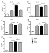
Analysis of cytokines following inhibition of canine PBMCs by Di-CPI. Supernatants of canine PBMCs stimulated for 48 h were subjected to ELISA to measure the concentration of IL-10, IL-12, TNF-α, IFN-γ, and IL-4. rDi-CPI treatment increased anti-inflammatory cytokine IL-10 (A), followed by significant decrease of the production of IL-12 (B) and TNF-α (C). IFN-γ (D) and IL-4 (E) showed no significant difference compared with controls. Naive indicates no protein was added to wells. Data are mean ± SEM of 3 samples with triplicate wells; representative data from one of three independent experiments. n.s., not significant. Statistically significant differences (* p < 0.05, ** p < 0.01) were determined by Mann–Whitney U-tests.

Similar articles

Cited by

References

    1. Simon F., Siles-Lucas M., Morchon R., Gonzalez-Miguel J., Mellado I., Carreton E., Montoya-Alonso J.A. Human and animal dirofilariasis: The emergence of a zoonotic mosaic. Clin. Microbiol Rev. 2012;25:507–544. doi: 10.1128/CMR.00012-12. - DOI - PMC - PubMed
    1. Bockova E., Iglodyova A., Kocisova A. Potential mosquito (Diptera:Culicidae) vector of Dirofilaria repens and Dirofilaria immitis in urban areas of Eastern Slovakia. Parasitol Res. 2015;114:4487–4492. doi: 10.1007/s00436-015-4692-8. - DOI - PubMed
    1. Dillon R.A. Influence of number of parasites and exercise on the severity of heartworm disease in dogs. Proc. Heartworm Symp. 1995;113:95.
    1. Kitagawa H., Ishihara K., Sasaki Y. Canine dirofilarial hemoglobinuria: Changes in right heart hemodynamics and heartworm migration from pulmonary artery towards right atrium following β 1-blocker administration. J. Vet. Med. Sci. 1987;49:1081–1086. doi: 10.1292/jvms1939.49.1081. - DOI - PubMed
    1. Simón F., Lopez-Belmonte J., Marcos-Atxutegi C., Morchón García R., R Martín-Pacho J. What is happening outside North America regarding human Dirofilariasis? Vet. Parasitol. 2005;133:181–189. doi: 10.1016/j.vetpar.2005.03.033. - DOI - PubMed

Publication types

MeSH terms

LinkOut - more resources