. 2019 Apr 13;11(4):531.
doi: 10.3390/cancers11040531.

Efficacy of a Selective Binder of αVβ3 Integrin Linked to the Tyrosine Kinase Inhibitor Sunitinib in Ovarian Carcinoma Preclinical Models

Affiliations

Efficacy of a Selective Binder of αVβ3 Integrin Linked to the Tyrosine Kinase Inhibitor Sunitinib in Ovarian Carcinoma Preclinical Models

Andrea Sartori et al. Cancers (Basel). .

Abstract

Ovarian carcinoma, the most lethal gynecological cancer, is characterized by late diagnosis, with drug resistance limiting the efficacy of platinum-based therapy. Since some integrins are upregulated in cancer, including ovarian carcinoma, they represent a potential target for drug delivery. Receptor tyrosine kinases are also deregulated in cancer and their expression has been associated with drug resistance. Here, the antitumor effects of three conjugates possessing a selective binder of the extracellular portion of integrin αVβ3 covalently linked to the tyrosine kinase inhibitor sunitinib were investigated in cisplatin-sensitive and -resistant ovarian carcinoma cells expressing both tyrosine kinase VEGFR2 and αVβ3 at different levels. We found that one of the three compounds was active in inhibiting the growth of both drug-sensitive and -resistant cells in the micromolar range with a slightly increased potency in resistant cells as compared to sunitinib. The same compound markedly impaired cell migratory and invasive abilities and reduced paxillin phosphorylation. Antitumor activity studies in IGROV-1/Pt1 cells xenografted in nude mice revealed a striking activity of this conjugate versus sunitinib. Taken together, our results support the interest of integrin-targeted sunitinib conjugates for the treatment of drug-resistant tumors.

Keywords: RGD ligands; RTK inhibitors; integrins; molecular conjugates; ovarian carcinoma; sunitinib.


Conflict of interest statement

The authors declare no conflict of interest.

Figures

Figure 1
Figure 1
Molecular structures of conjugates 13 and the parent sunitinib and c(AmpRGD) components.
Figure 2
Figure 2
Western blot analysis of VEGFR2 levels in different ovarian carcinoma cell lines. Western blot analysis was carried out in ovarian carcinoma exponentially growing cells. Control loading is shown by vinculin. One experiment representative of three is reported. For whole Western blot analysis of VEGFR2 levels and band intensities quantification of VEGFR2 levels in different ovarian carcinoma cell lines, see Figure S3 and Table S1, respectively.
Figure 3
Figure 3
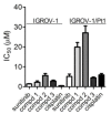
Sensitivity to sunitinib, conjugates 13 and cisplatin as assessed by cell growth inhibition assays. Cells were seeded and 24 h later exposed to the compounds for 72 h. Cells were then counted using a cell counter. IC50 is defined as the concentration inhibiting cell growth by 50%. The IC50 values of the c(AmpRGD) per se were 6.04 ± 2.8 μM in IGROV-1 cells and >30 μM in IGROV-1/Pt1 cells. Sensitivity relative to sunitinib is: 1.59, 4.00, 2.01 for compounds 1, 2, and 3, respectively, in IGROV-1 cells, and 3.88, 5.28, 0.86 for compounds 1, 2, and 3, respectively, in IGROV-1/Pt1 cells.
Figure 4
Figure 4
Modulation of migratory and invasive abilities of IGROV-1 and IGROV-1/Pt1 cells by sunitinib and selective binders of αVβ3 integrin namely, c(AmpRGD) alone or linked to sunitinib (compounds 13). Cells were subjected to migration (a) and invasion assays (b) in serum-free medium using transwell plates after exposure to the compounds (10 μM). Migrating and invading cells were counted under a light microscope. Columns represent cell numbers/field (± SE; n = 3).
Figure 5
Figure 5
Western blot analysis of paxillin and phosphorylated-paxillin (p-paxillin) levels in IGROV-1 and IGROV-1/Pt1 cells. Cells were exposed for 24 h to sunitinib or to selective binders of αVβ3 integrin linked to sunitinib (compounds 13) at the indicated concentrations and then harvested for western blot analysis. Tubulin was used as loading control. A representative experiment is shown (for quantification of band intensities, see Table S2). For whole Western blot analysis of paxillin and phospho-paxillin levels in IGROV-1 and IGROV-1/Pt1 cells, see Figure S4.
Figure 6
Figure 6
Antitumor activity of conjugate 3 in IGROV-1/Pt1 tumors. The effects of i.p. compound 3, 20 mg/kg (having a sunitinib-equivalent loading 9 times lower than parental sunitinib), oral sunitinib, 40 mg/kg, qd×5/w×4w and intravenous cisplatin (4.5 mg/Kg, q7d×3) were evaluated on the growth of the IGROV-1/Pt1 carcinoma cells s.c. injected into the right flank of female nude mice (107 cells/mouse) on day 0. Treatment started on day 4: compound 3 (open up triangle), sunitinib (filled square), cisplatin (down triangle) or control (open circle). Experimental groups consisted of seven animals. Growth curves of the tumors are shown. Points report mean values of tumor volumes. Bars: SD.
Figure 7
Figure 7
Evaluation of microvessels after treatment. Histopathological analysis of IGROV-1/Pt1 xenograft samples after treatment with compound 3 or sunitinib. (a) Quantitative analysis of tumor associated vasculature. CD31-positive area was measured in three randomly selected 200× microscopic fields from seven samples per group. The reported numbers correspond to the mean of CD31 positive areas in analyzed groups. Tumors were obtained from mice sacrificed 24 h after the last treatment; *** correspond to p < 0.001 by One Way ANOVA followed by Bonferroni Multiple Comparison test. (b) Representative 200× microscopic fields from a control group sample, and tumor from a sample treated with compound 3 and sunitinib. The scale bar corresponds to 100 μm.

Similar articles

Cited by

References

    1. Siegel R.L., Miller K.D., Jemal A. Cancer statistics, 2018. CA Cancer. J. Clin. 2018;68:7–30. doi: 10.3322/caac.21442. - DOI - PubMed
    1. Bast R.C., Jr., Hennessy B., Mills G.B. The biology of Ovarian cancer: New opportunities for translation. Nat. Rev. Cancer. 2009;9:415–428. doi: 10.1038/nrc2644. - DOI - PMC - PubMed
    1. Cannistra S.A. Cancer of the ovary. N. Engl. J. Med. 2004;351:2519–2529. doi: 10.1056/NEJMra041842. - DOI - PubMed
    1. Kelland L. The Resurgence of platinum-based cancer chemotherapy. Nat. Rev. Cancer. 2007;7:573–584. doi: 10.1038/nrc2167. - DOI - PubMed
    1. Cossa G., Gatti L., Zunino F., Perego P. Strategies to improve the efficacy of platinum compounds. Curr. Med. Chem. 2009;16:2355–2365. doi: 10.2174/092986709788682083. - DOI - PubMed