. 2019 Apr 16;20(8):1873.
doi: 10.3390/ijms20081873.

The Histone Deacetylase Inhibitor Suberoylanilide Hydroxamic Acid (SAHA) Restores Cardiomyocyte Contractility in a Rat Model of Early Diabetes

Affiliations

The Histone Deacetylase Inhibitor Suberoylanilide Hydroxamic Acid (SAHA) Restores Cardiomyocyte Contractility in a Rat Model of Early Diabetes

Leonardo Bocchi et al. Int J Mol Sci. .

Abstract

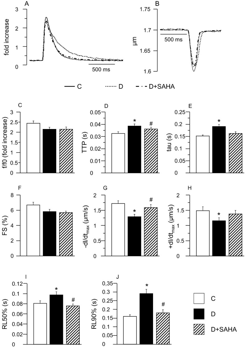
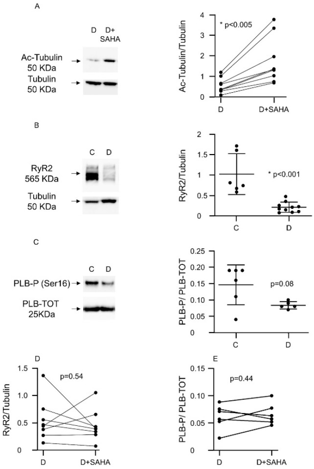
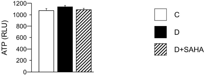
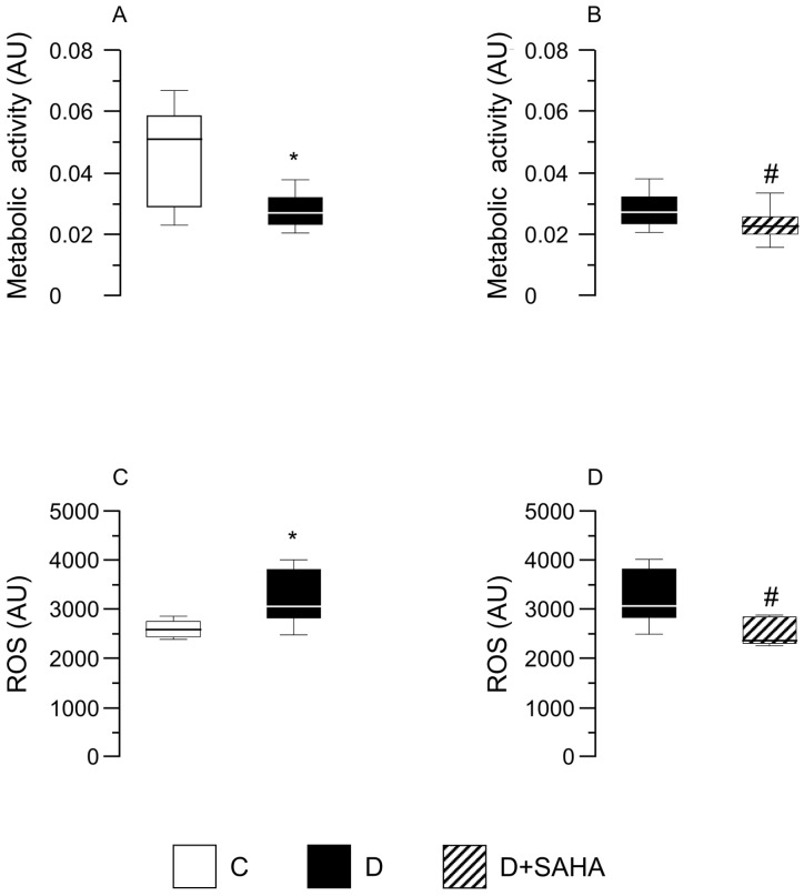
In early diabetes, hyperglycemia and the associated metabolic dysregulation promote early changes in the functional properties of cardiomyocytes, progressively leading to the appearance of the diabetic cardiomyopathy phenotype. Recently, the interplay between histone acetyltransferases (HAT) and histone deacetylases (HDAC) has emerged as a crucial factor in the development of cardiac disorders. The present study evaluates whether HDAC inhibition can prevent the development of cardiomyocyte contractile dysfunction induced by a short period of hyperglycemia, with focus on the potential underlying mechanisms. Cell contractility and calcium dynamics were measured in unloaded ventricular myocytes isolated from the heart of control and diabetic rats. Cardiomyocytes were either untreated or exposed to the pan-HDAC inhibitor suberoylanilide hydroxamic acid (SAHA) for 90 min. Then, a fraction of each group of cells was used to evaluate the expression levels of proteins involved in the excitation-contraction coupling, and the cardiomyocyte metabolic activity, ATP content, and reactive oxygen species levels. SAHA treatment was able to counteract the initial functional derangement in cardiomyocytes by reducing cell oxidative damage. These findings suggest that early HDAC inhibition could be a promising adjuvant approach for preventing diabetes-induced cardiomyocyte oxidative damage, which triggers the pro-inflammatory signal cascade, mitochondrial damage, and ventricular dysfunction.

Keywords: HDAC inhibition; calcium transients; cardiomyocyte mechanics; cell oxidative stress; diabetes.


Conflict of interest statement

The authors declare no conflict of interest. The funders had no role in the design of the study; in the collection, analyses, or interpretation of data; in the writing of the manuscript, or in the decision to publish the results.

Figures

Figure 1
Figure 1
Effect of suberoylanilide hydroxamic acid (SAHA) treatment on calcium transients and cell mechanics in left ventricular myocytes. (A,B) Representative examples of calcium transients (A: normalized traces) and sarcomere shortening (B) recorded from control (C, solid line), diabetic (D, dotted line) and SAHA treated diabetic cells (D + SAHA, dashed line). In bar graphs CJ: Mean values ± SEM of the amplitude of calcium transient (C: f/f0), time-to-peak of the calcium transient (D: TTP), time constant of the rate of intracellular Ca2+ clearing (E: tau), fraction of shortening (F: FS), maximal rate of shortening (G: −dL/dtmax), maximal rate of re-lengthening (H, +dL/dtmax), time to 50% and 90% re-lengthening (I: RL50%; J: RL90%), measured in 45 C, 61 D, and 48 D + SAHA cardiomyocytes (CMs). * p < 0.05 significant differences vs. C; # p < 0.05 significant differences vs. D. (GLM-ANOVA for repeated measurements).
Figure 2
Figure 2
Effect of SAHA treatment on protein expression in left ventricular myocytes. (A) Western blot panels and densitometric analysis showing Ac-Tubulin protein expression after SAHA treatment in ventricular CMs isolated from nine D rats. Wilcoxon matched-pairs signed rank test: * p < 0.05 vs. C; (B) Western blot panels and densitometric analysis showing RyR2 protein expression reduction in CMs of diabetic rats (n. of rats = 10) compared to C cells (n. of rats = 6). Mann–Whitney U-test: * p < 0.001 vs. C; (C) Western blot panels and densitometric analysis showing the expression of phosphorylated phospholamban (PLB-P, Ser16) compared to total phospholamban (PLB-TOT), in adult C cardiomyocytes vs. D cells (n. of C rats = 6; n. of D rats = 5), normalized to total protein concentration (Ponceau). Mann–Whitney U-test: p = 0.08 vs. C; (D) Western blot panels and densitometric analysis showing RyR2 protein expression after SAHA treatment in diabetic CMs (n. of rats = 8). Wilcoxon matched-pairs signed rank test; (E) Western blot analysis showing the expression of phosphorylated phospholamban (PLB-P, Ser16) compared to total phospholamban (PLB-TOT) in adult rat CMs after SAHA treatment normalized to total protein concentration (Ponceau) (n. of rats = 6). Wilcoxon matched-pairs signed rank test.
Figure 3
Figure 3
Effect of SAHA treatment on cardiomyocyte ATP content. Mean values ± SEM of ATP content in control (C), diabetic (D), and SAHA-treated D cells (D + SAHA). Values are expressed as relative light units (RLU) counts/s. Kruskal–Wallis and U Mann–Whitney test.
Figure 4
Figure 4
Effect of SAHA treatment on CMs’ metabolic activity and intracellular ROS content. Data are reported as median values and interquartile range. In left panels: Effect of diabetes on CM metabolic activity (A) and ROS levels (C), compared with control cells. Mann–Whitney U-test: * p < 0.05 vs. group C. In right panels: metabolic activity (B) and ROS content (D) measured in diabetic CMs before and after SAHA-exposure. Cells were isolated from the heart of three normal rats (group C) and five diabetic rats (groups D and D + SAHA). AU = arbitrary units. # p < 0.05 vs. D group; Wilcoxon matched-pairs signed rank test.

Similar articles

Cited by

References

    1. Hölscher M.E., Bode C., Bugger H. Diabetic Cardiomyopathy: Does the Type of Diabetes Matter? Int. J. Mol. Sci. 2016;17:2136. doi: 10.3390/ijms17122136. - DOI - PMC - PubMed
    1. Zhang L., Du J., Yano N., Wang H., Zhao Y.T., Dubielecka-Szczerba P., Zhuang S., Chin E.Y., Qin G., Zhao T.C. Sodium butyrate protects against high fat diet-induced cardiac dysfunction and metabolic disorders in type II diabetic mice. J. Cell Biochem. 2017;118:2395–2408. doi: 10.1002/jcb.25902. - DOI - PMC - PubMed
    1. Jia G., Whaley-Connell A., Sowers J.R. Diabetic cardiomyopathy: A hyperglycaemia- and insulin-resistance-induced heart disease. Diabetologia. 2018;61:21–28. doi: 10.1007/s00125-017-4390-4. - DOI - PMC - PubMed
    1. Savi M., Bocchi L., Sala R., Frati C., Lagrasta C., Madeddu D., Falco A., Pollino S., Bresciani L., Miragoli M., et al. Parenchymal and Stromal Cells Contribute to Pro-Inflammatory Myocardial Environment at Early Stages of Diabetes: Protective Role of Resveratrol. Nutrients. 2016;8:729. doi: 10.3390/nu8110729. - DOI - PMC - PubMed
    1. Bugger H., Abel E.D. Molecular mechanisms of diabetic cardiomyopathy. Diabetologia. 2014;57:660–671. doi: 10.1007/s00125-014-3171-6. - DOI - PMC - PubMed

LinkOut - more resources