

Efficient gene transfer into T lymphocytes by fiber-modified human adenovirus 5
- PMID: 31014302
- PMCID: PMC6480437
- DOI: 10.1186/s12896-019-0514-x
Efficient gene transfer into T lymphocytes by fiber-modified human adenovirus 5
Abstract
Background: The gene transduction efficiency of adenovirus to hematopoietic cells, especially T lymphocytes, is needed to be improved. The purpose of this study is to improve the transduction efficiency of T lymphocytes by using fiber-modified human adenovirus 5 (HAdV-5) vectors.
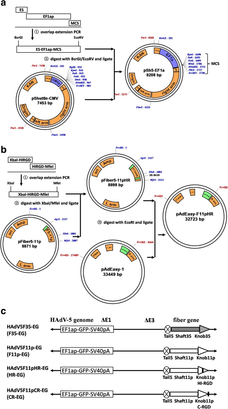
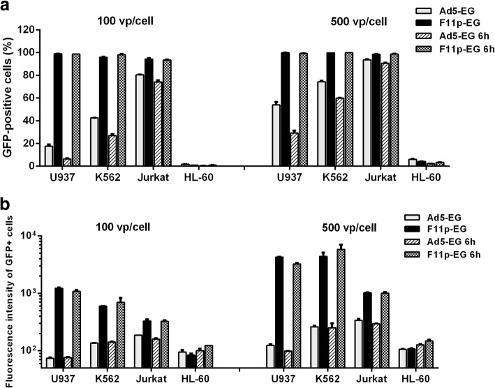
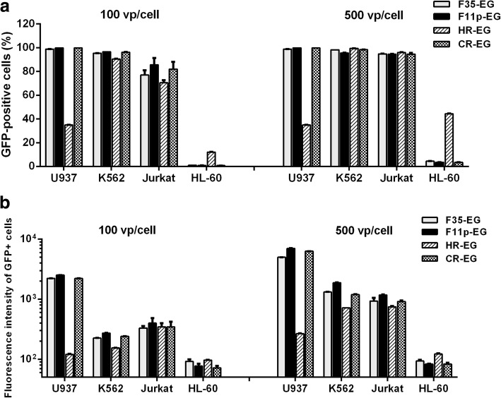
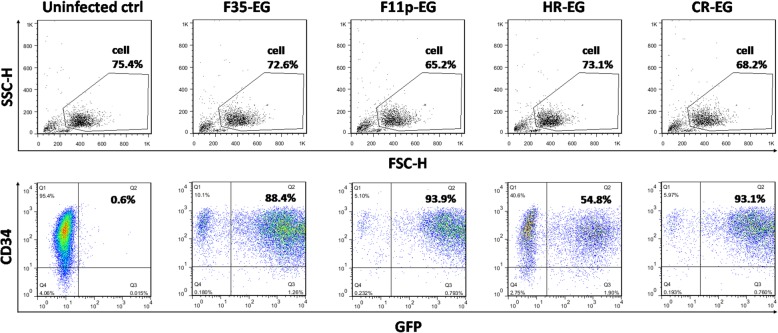
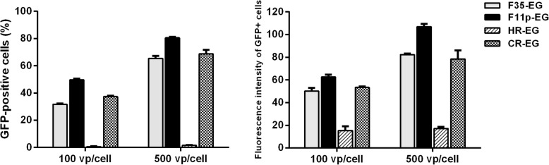
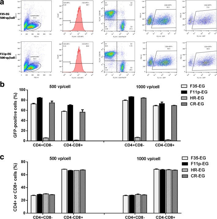
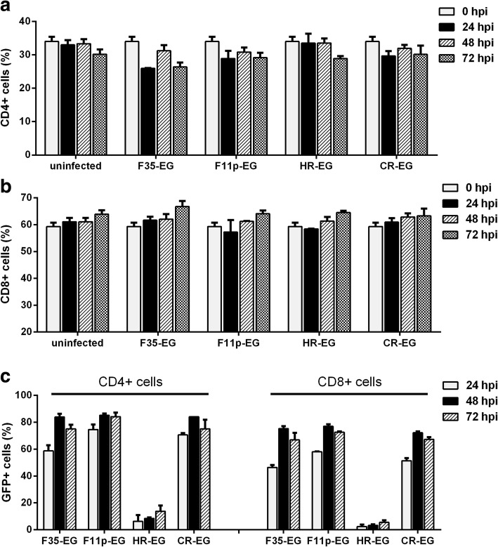
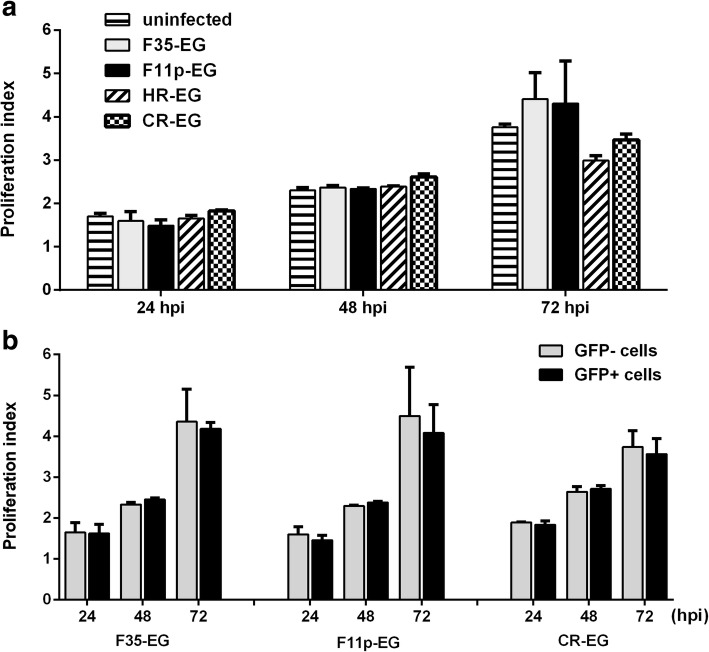
Results: Four fiber-modified human adenovirus 5 (HAdV-5) vectors were investigated to transduce hematopoietic cells. F35-EG or F11p-EG were HAdV-35 or HAdV-11p fiber pseudotyped HAdV-5, and HR-EG or CR-EG vectors were generated by incorporating RGD motif to the HI loop or to the C-terminus of F11p-EG fiber. All vectors could transduce more than 90% of K562 or Jurkat cells at an multiplicity of infection (MOI) of 500 viral particle per cell (vp/cell). All vectors except HR-EG could transduce nearly 90% cord blood CD34+ cells or 80% primary human T cells at the MOI of 1000, and F11p-EG showed slight superiority to F35-EG and CR-EG. Adenoviral vectors transduced CD4+ T cells a little more efficiently than they did to CD8+ T cells. These vectors showed no cytotoxicity at an MOI as high as 1000 vp/cell because the infected and uninfected T cells retained the same CD4/CD8 ratio and cell growth rate.
Conclusions: HAdV-11p fiber pseudotyped HAdV-5 could effectively transduce human T cells when human EF1a promoter was used to control the expression of transgene, suggesting its possible application in T cell immunocellular therapy.
Keywords: Adenovirus 5 vector; Gene therapy; Human hematopoietic cells.
Conflict of interest statement
Ethics approval and consent to participate
Because the ways to get samples have no effect on the health of donors and the samples are not for clinical use, the local ethics committee (Medical ethics committee of affiliated hospital of Qingdao university) agreed to the protocols (Hematopoietic Cell Transduction 2018–03) of this project in which oral inform consent is needed. All donors are oral informed in conversation room under video record.
Consent for publication
Not applicable.
Competing interests
The authors declare that they have no competing interests.
Publisher’s Note
Springer Nature remains neutral with regard to jurisdictional claims in published maps and institutional affiliations.
Figures
Similar articles
-
Fiber modifications enable fowl adenovirus 4 vectors to transduce human cells.J Gene Med. 2021 Oct;23(10):e3368. doi: 10.1002/jgm.3368. Epub 2021 Jun 11. J Gene Med. 2021. PMID: 34050587 Free PMC article.
-
Gene transfer into human T lymphocytes and natural killer cells by Ad5/F35 chimeric adenoviral vectors.Exp Hematol. 2004 Jun;32(6):536-46. doi: 10.1016/j.exphem.2004.03.010. Exp Hematol. 2004. PMID: 15183894
-
Development of a novel adenovirus-alphavirus hybrid vector with RNA replicon features for malignant hematopoietic cell transduction.Cancer Gene Ther. 2013 Aug;20(8):429-36. doi: 10.1038/cgt.2013.37. Epub 2013 Jul 5. Cancer Gene Ther. 2013. PMID: 23827880
-
Tropism and transduction of oncolytic adenovirus 5 vectors in cancer therapy: Focus on fiber chimerism and mosaicism, hexon and pIX.Virus Res. 2018 Sep 15;257:40-51. doi: 10.1016/j.virusres.2018.08.012. Epub 2018 Aug 17. Virus Res. 2018. PMID: 30125593 Review.
-
The importance of coagulation factors binding to adenovirus: historical perspectives and implications for gene delivery.Expert Opin Drug Deliv. 2014 Nov;11(11):1795-813. doi: 10.1517/17425247.2014.938637. Epub 2014 Jul 18. Expert Opin Drug Deliv. 2014. PMID: 25036189 Review.
Cited by
-
Fiber modifications enable fowl adenovirus 4 vectors to transduce human cells.J Gene Med. 2021 Oct;23(10):e3368. doi: 10.1002/jgm.3368. Epub 2021 Jun 11. J Gene Med. 2021. PMID: 34050587 Free PMC article.
-
Superior infectivity of the fiber chimeric oncolytic adenoviruses Ad5/35 and Ad5/3 over Ad5-delta-24-RGD in primary glioma cultures.Mol Ther Oncolytics. 2021 Dec 21;24:230-248. doi: 10.1016/j.omto.2021.12.013. eCollection 2022 Mar 17. Mol Ther Oncolytics. 2021. PMID: 35071746 Free PMC article.
-
Restriction-Assembly: A Solution to Construct Novel Adenovirus Vector.Viruses. 2022 Mar 6;14(3):546. doi: 10.3390/v14030546. Viruses. 2022. PMID: 35336953 Free PMC article.
-
Bovine Adenovirus-3 Tropism for Bovine Leukocyte Sub-Populations.Viruses. 2020 Dec 12;12(12):1431. doi: 10.3390/v12121431. Viruses. 2020. PMID: 33322850 Free PMC article.
References
Publication types
MeSH terms
Substances
LinkOut - more resources
Full Text Sources
Other Literature Sources
Research Materials
Miscellaneous
