Review
. 2019 Apr 23;26(1):28.
doi: 10.1186/s12929-019-0520-2.

Molecular mechanisms in progression of HPV-associated cervical carcinogenesis

Affiliations
Review

Molecular mechanisms in progression of HPV-associated cervical carcinogenesis

Sadhana M Gupta et al. J Biomed Sci. .

Retraction in

Abstract

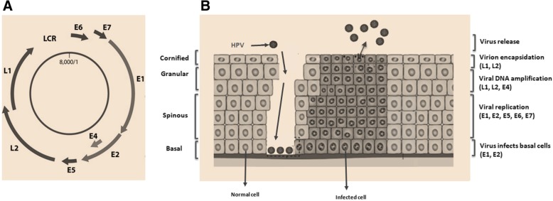
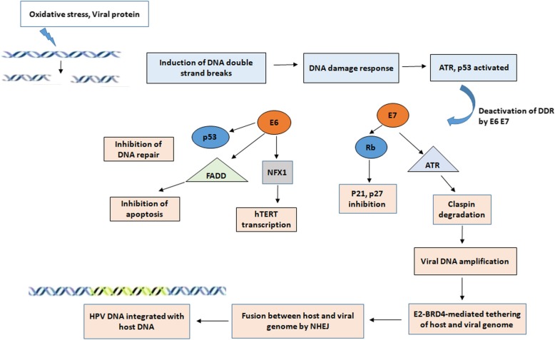
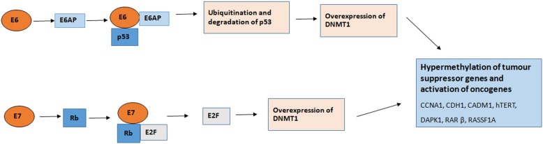
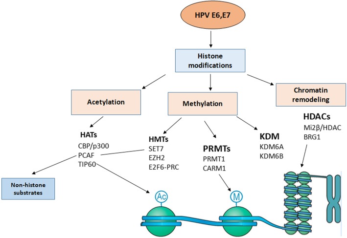
Cervical cancer is the fourth most frequent cancer in women worldwide and a major cause of mortality in developing countries. Persistent infection with high-risk human papillomavirus (HPV) is a necessary cause for the development of cervical cancer. In addition, genetic and epigenetic alterations in host cell genes are crucial for progression of cervical precancerous lesions to invasive cancer. Although much progress has been made in understanding the life cycle of HPV and it's role in the development of cervical cancer, there is still a critical need for accurate surveillance strategies and targeted therapeutic options to eradicate these cancers in patients. Given the widespread nature of HPV infection and the type specificity of currently available HPV vaccines, it is crucial that molecular details of the natural history of HPV infection as well as the biological activities of viral oncoproteins be elucidated. A better understanding of the mechanisms involved in oncogenesis can provide novel insights and opportunities for designing effective therapeutic approaches against HPV-associated malignancies. In this review, we briefly summarize epigenetic alterations and events that cause alterations in host genomes inducing cell cycle deregulation, aberrant proliferation and genomic instability contributing to tumorigenesis.

Keywords: Cervical carcinogenesis; Chromosomal mutations; Epigenetic alterations; Oncogene expression.


Conflict of interest statement

Authors’ information

None.

Ethics approval and consent to participate

Not applicable (The present paper does not report on or involve the use of any animal or human data or tissue).

Consent for publication

Not applicable (The present paper does not contain data from any individual person).

Competing interests

The authors declare that they have no competing interests.

Publisher’s Note

Springer Nature remains neutral with regard to jurisdictional claims in published maps and institutional affiliations.

Figures

Fig. 1
Fig. 1
Organization of HPV genome. a. HPV genome has a circular double-stranded DNA (8000bp). The viral genes are transcribed in a single direction (clockwise). There are genes coding for non-structural proteins (E1, E2, E4, E5, E6, and E7) and structural proteins (L1, L2), and a transcriptional control region (long control region; LCR). LCR contains a DNA replication origin and functions as a regulator for DNA replication. b. The HPV lifecycle. Human papillomavirus is thought to reach the basal cells through microabrasions in the cervical epithelium. After infection, the early human papillomavirus genes E1, E2, E4, E5, E6, and E7 are expressed and the viral DNA replicates from episomal DNA and establishes ∼50 HPV episome copies, which then segregate between the daughter progeny as the cells undergo cell division. In the upper layers of epithelium, viral genome is replicated further, and late genes L1 and L2, and E4 are expressed. The early viral proteins E6 and E7 are key to stimulating proliferation and milieu for E1 and E2-driven viral genome replication to a high copy number. Integration of human papillomavirus genome into the host chromosome occurs with associated loss or disruption of E2, and subsequent upregulation of E6 and E7 oncogene expression. Terminal differentiation of infected cells in the upper epithelial layers activates the expression of E4 and then L1 and L2 to encapsidate the viral genomes to form progeny virions. The shed virus can then initiate a new infection
Fig. 2
Fig. 2
Mechanism of HPV DNA integration: Induction of double strand breaks by reactive oxygen species, viral protein induces DNA damage response, following which ATR and P53 get activated to repair the damage. HPV oncogenes deactivate the normal function of DNA damage response (DDR). E7 acts on ATR and degrades claspin, inactivates Rb disrupting the cell cycle inhibitors p21, p27. E6 degrades p53 inhibiting DNA repair, degrades FAS-associated death domain protein (FADD) preventing apoptosis via Fas pathway, promotes degradation of transcriptional repressor, NFX1 and activates hTERT transcription. Virus utilizes the DDR machinery for its replication and close proximity with host genome is mediated by the Bromodomain protein 4, BRD4-E2 complex. The fusion between host and viral genome is accomplished by Nonhomologous mediated end joining (NHEJ) finally leading to integration of HPV DNA into the host genome
Fig. 3
Fig. 3
Schematic presentation of DNA methylation by E6 and E7 oncoproteins: Binding of E6-E6AP complex to p53 to leads to ubiquitination and degradation of p53 and induces overexpression and activity of DNA methyltransferase (DNMT) 1. Binding of E7 to pRb causes the release of E2F, favoring the overexpression of DNMT1. The activities of E6 and E7 causing increase in activity of DNMT1 results in hypermethylation of tumor-suppressor gene promoters, leading to silencing of genes, cellular transformation and tumorigenesis
Fig. 4
Fig. 4
Schematic presentation of Histone modifications by HPV E6 and E7 interaction with cellular epigenetic modifiers. HPV oncoproteins E6, E7 bind to and/or modulate the expression of histone modifying enzymes, class I Histone deacetylases (HDAC), Histone acetyltransferases (HATs), Histone lysine demethylases (KDMs) and subunits of chromatin remodeling complexes. These interactions contribute to chromatin remodelling and transcriptional regulation leading to either activation or repression of gene expression
Fig. 5
Fig. 5
Schematic structure of oncoprotein E6. Protein structure and functions of HPV16 E6. Four zinc-binding motifs are indicated as grey boxes. The two zinc fingers are shown together with regions that are involved in interacting with some of its cellular target proteins. E6 contains PDZ domain–binding motif at its C-terminal extremity and three Nuclear localization signals (NLS). Functions associated with proteins in different regions are indicated by arrows
Fig. 6
Fig. 6
Schematic structure of oncoprotein E7. Protein structure and functions of HPV16 E7 and the most important amino acid motifs required for integrity and protein functions. Relative locations of the regions with sequence motifs similar to a portion of conserved region 1 (CR1) and the entire CR2 of adenovirus E1A are shown with the pRB-binding site LXCXE in the CR2. Zinc binding motifs are indicated in grey boxes. The zinc finger is shown together with the regions involved in pRb binding (LXCXE) and the two serine residues (31 and 32) that are susceptible to casein kinase II (CKII) phosphorylation. Functions associated with proteins in different regions are indicated by PI3K
Fig. 7
Fig. 7
Molecular events in progression to cervical carcinogenesis. Persistent infection with high risk HPV leads to integration of HPV into the host genome, leading to overexpression of oncogenes E6 And E7. Interaction of Е7 with pRb protein leads to aberrant initiation of S-phase, release of E2F transcription factor that triggers the expression of cyclins and CDK inhibitors p21 and p27, altering the integrity of cell cycle thereby contributing to cellular immortalization and transformation. Е6 targets р53 for proteasomal degradation leading to inhibition of apoptosis and DNA repair. E6 activates PI3K/Akt pathway, interacts with cellular proteins NFX1 and induces activation of hTERT leading to immortalization and transformation. The interaction of both onoproteins with DNMTs leads to aberrant methylation causing silencing of tumor suppressor genes. E7 interaction with HDACs causes chromosome remodeling and genome instability. Thus, the cross interaction of E6 and E7 with various pathways plays a key role in progression to carcinogenesis

Similar articles

Cited by

References

    1. Dunne EF, Unger ER, Sternberg M, McQuillan G, Swan DC, Patel SS, Markowitz LE. Prevalence of HPV infection among females in the United States. JAMA. 2007;297:813–819. doi: 10.1001/jama.297.8.813. - DOI - PubMed
    1. Laudadio J. Human papillomavirus detection: testing methodologies and their clinical utility in cervical cancer screening. Adv Anat Pathol. 2013;20:158–167. doi: 10.1097/PAP.0b013e31828d1893. - DOI - PubMed
    1. Rodríguez AC, Schiffman M, Herrero R, Wacholder S, Hildesheim A, Castle PE, Solomon D, Burk R. Rapid clearance of human papillomavirus and implications for clinical focus on persistent infections. J Natl Cancer Inst. 2008;100:513–517. doi: 10.1093/jnci/djn044. - DOI - PMC - PubMed
    1. Castle PE, Fetterman B, Poitras N, Lorey T, Shaber R, Kinney W. Five-year experience of human papillomavirus DNA and Papanicolaou test cotesting. Obstet Gynecol. 2009;113:595–600. doi: 10.1097/AOG.0b013e3181996ffa. - DOI - PMC - PubMed
    1. Cubie HA, Seagar AL, Beattie GJ, Monaghan S, Williams AR. A longitudinal study of HPV detection and cervical pathology in HIV infected women. Sex Transm Infect. 2000;76:257–261. doi: 10.1136/sti.76.4.257. - DOI - PMC - PubMed