

STIM1 at the plasma membrane as a new target in progressive chronic lymphocytic leukemia
- PMID: 31014395
- PMCID: PMC6480884
- DOI: 10.1186/s40425-019-0591-3
STIM1 at the plasma membrane as a new target in progressive chronic lymphocytic leukemia
Abstract
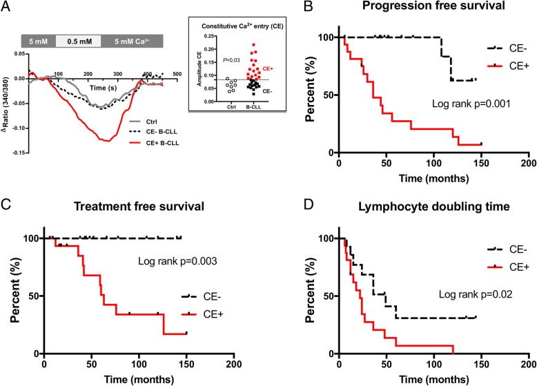
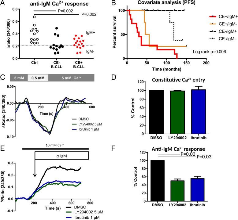
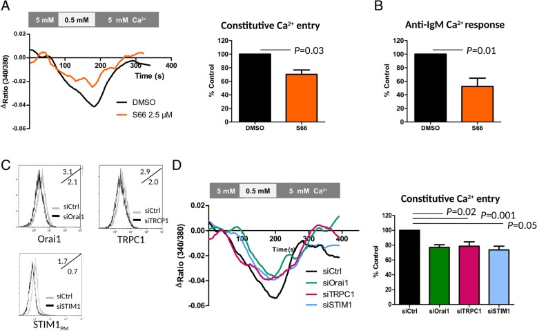
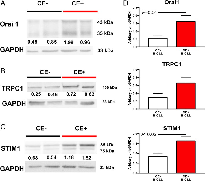
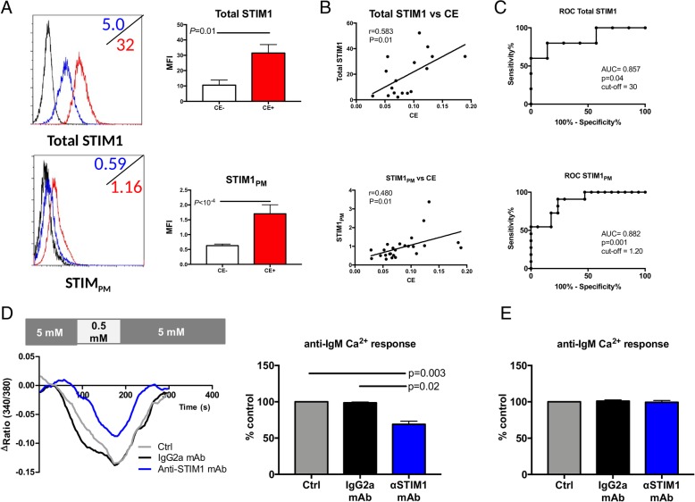
Background: Dysregulation in calcium (Ca2+) signaling is a hallmark of chronic lymphocytic leukemia (CLL). While the role of the B cell receptor (BCR) Ca2+ pathway has been associated with disease progression, the importance of the newly described constitutive Ca2+ entry (CE) pathway is less clear. In addition, we hypothesized that these differences reflect modifications of the CE pathway and Ca2+ actors such as Orai1, transient receptor potential canonical (TRPC) 1, and stromal interaction molecule 1 (STIM1), the latter being the focus of this study.
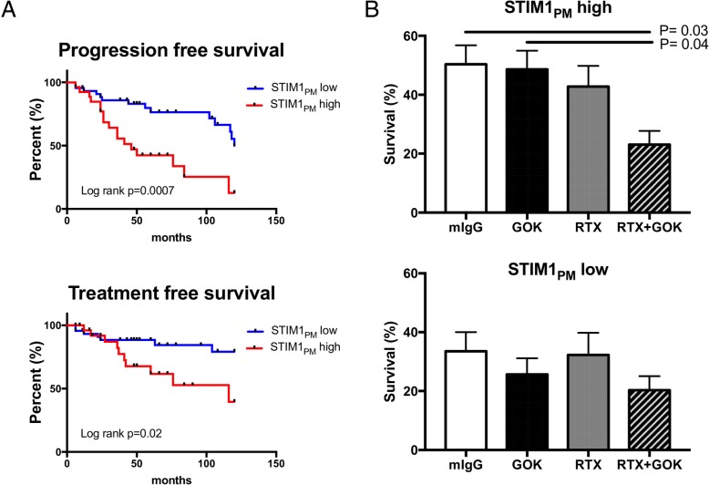
Methods: An extensive analysis of the Ca2+ entry (CE) pathway in CLL B cells was performed including constitutive Ca2+ entry, basal Ca2+ levels, and store operated Ca2+ entry (SOCE) activated following B cell receptor engagement or using Thapsigargin. The molecular characterization of the calcium channels Orai1 and TRPC1 and to their partner STIM1 was performed by flow cytometry and/or Western blotting. Specific siRNAs for Orai1, TRPC1 and STIM1 plus the Orai1 channel blocker Synta66 were used. CLL B cell viability was tested in the presence of an anti-STIM1 monoclonal antibody (mAb, clone GOK) coupled or not with an anti-CD20 mAb, rituximab. The Cox regression model was used to determine the optimal threshold and to stratify patients.
Results: Seeking to explore the CE pathway, we found in untreated CLL patients that an abnormal CE pathway was (i) highly associated with the disease outcome; (ii) positively correlated with basal Ca2+ concentrations; (iii) independent from the BCR-PLCγ2-InsP3R (SOCE) Ca2+ signaling pathway; (iv) supported by Orai1 and TRPC1 channels; (v) regulated by the pool of STIM1 located in the plasma membrane (STIM1PM); and (vi) blocked when using a mAb targeting STIM1PM. Next, we further established an association between an elevated expression of STIM1PM and clinical outcome. In addition, combining an anti-STIM1 mAb with rituximab significantly reduced in vitro CLL B cell viability within the high STIM1PM CLL subgroup.
Conclusions: These data establish the critical role of a newly discovered BCR independent Ca2+ entry in CLL evolution, provide new insights into CLL pathophysiology, and support innovative therapeutic perspectives such as targeting STIM1 located at the plasma membrane.
Trial registration: ClinicalTrials.gov NCT03294980.
Keywords: CLL; Constitutive Ca2+ entry; Disease outcome; STIM1.
Conflict of interest statement
Ethics approval and consent to participate
This study was approved by the Ethical Board at the Brest University Hospital (OFICE, November 26th, 2015; clinicaltrials: NCT03294980) in accordance with the declaration of Helsinki.
Consent for publication
Written informed consent was obtained from all of the patients.
Competing interests
The authors declare that they have no competing interests.
Publisher’s Note
Springer Nature remains neutral with regard to jurisdictional claims in published maps and institutional affiliations.
Figures
Similar articles
-
Local Ca²+ entry via Orai1 regulates plasma membrane recruitment of TRPC1 and controls cytosolic Ca²+ signals required for specific cell functions.PLoS Biol. 2011 Mar;9(3):e1001025. doi: 10.1371/journal.pbio.1001025. Epub 2011 Mar 8. PLoS Biol. 2011. PMID: 21408196 Free PMC article.
-
Orai1 and Stim1 Mediate the Majority of Store-Operated Calcium Entry in Multiple Myeloma and Have Strong Implications for Adverse Prognosis.Cell Physiol Biochem. 2018;48(6):2273-2285. doi: 10.1159/000492645. Epub 2018 Aug 16. Cell Physiol Biochem. 2018. PMID: 30114708
-
STIM-TRP Pathways and Microdomain Organization: Contribution of TRPC1 in Store-Operated Ca2+ Entry: Impact on Ca2+ Signaling and Cell Function.Adv Exp Med Biol. 2017;993:159-188. doi: 10.1007/978-3-319-57732-6_9. Adv Exp Med Biol. 2017. PMID: 28900914 Review.
-
SARAF modulates TRPC1, but not TRPC6, channel function in a STIM1-independent manner.Biochem J. 2016 Oct 15;473(20):3581-3595. doi: 10.1042/BCJ20160348. Epub 2016 Aug 9. Biochem J. 2016. PMID: 27506849
-
TRPC1, Orai1, and STIM1 in SOCE: Friends in tight spaces.Cell Calcium. 2017 May;63:33-39. doi: 10.1016/j.ceca.2016.12.009. Epub 2016 Dec 30. Cell Calcium. 2017. PMID: 28089266 Free PMC article. Review.
Cited by
-
Defective interaction of mutant calreticulin and SOCE in megakaryocytes from patients with myeloproliferative neoplasms.Blood. 2020 Jan 9;135(2):133-144. doi: 10.1182/blood.2019001103. Blood. 2020. PMID: 31697806 Free PMC article.
-
Transient Receptor Potential (TRP) Channels in Haematological Malignancies: An Update.Biomolecules. 2021 May 20;11(5):765. doi: 10.3390/biom11050765. Biomolecules. 2021. PMID: 34065398 Free PMC article. Review.
-
Targeting metabolic reprogramming in chronic lymphocytic leukemia.Exp Hematol Oncol. 2022 Jun 27;11(1):39. doi: 10.1186/s40164-022-00292-z. Exp Hematol Oncol. 2022. PMID: 35761419 Free PMC article. Review.
-
Epigenetic Modifications in Generalized Autoimmune Epithelitis: Sjögren's Syndrome and Primary Biliary Cholangitis.Epigenomes. 2019 Aug 8;3(3):15. doi: 10.3390/epigenomes3030015. Epigenomes. 2019. PMID: 34968227 Free PMC article. Review.
-
Proton-sensing ion channels, GPCRs and calcium signaling regulated by them: implications for cancer.Front Cell Dev Biol. 2024 Mar 5;12:1326231. doi: 10.3389/fcell.2024.1326231. eCollection 2024. Front Cell Dev Biol. 2024. PMID: 38505262 Free PMC article. Review.
References
Publication types
MeSH terms
Substances
Associated data
LinkOut - more resources
Full Text Sources
Other Literature Sources
Medical
Miscellaneous