

Pericytes Act as Key Players in Spinal Cord Injury
- PMID: 31014955
- PMCID: PMC6717911
- DOI: 10.1016/j.ajpath.2019.03.008
Pericytes Act as Key Players in Spinal Cord Injury
Abstract
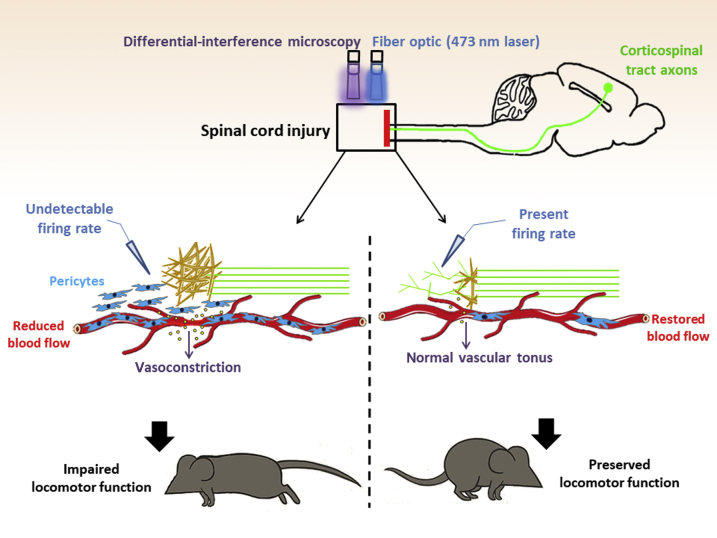
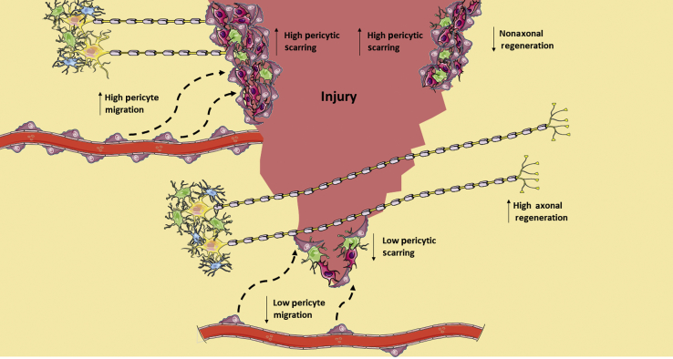
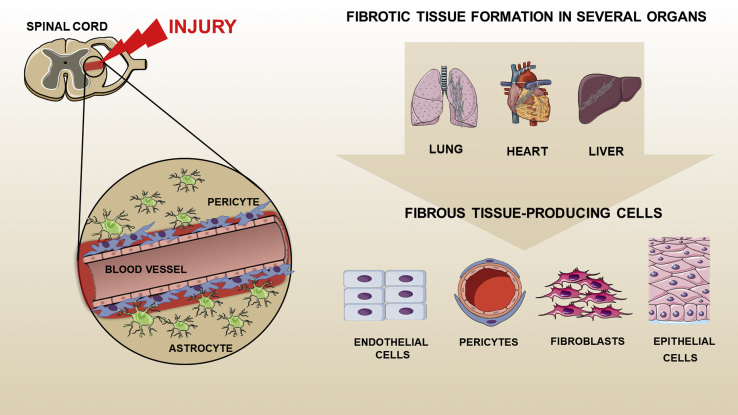
Spinal cord injury results in locomotor impairment attributable to the formation of an inhibitory fibrous scar, which prevents axonal regeneration after trauma. The scarcity of knowledge about the molecular and cellular mechanisms involved in scar formation after spinal cord lesion impede the design of effective therapies. Recent studies, by using state-of-the-art technologies, including genetic tracking and blockage of pericytes in combination with optogenetics, reveal that pericyte blockage facilitates axonal regeneration and neuronal integration into the local neural circuitry. Strikingly, a pericyte subset is essential during scarring after spinal cord injury, and its arrest results in motor performance improvement. The arising knowledge from current research will contribute to novel approaches to develop therapies for spinal cord injury. We review novel advances in our understanding of pericyte biology in the spinal cord.
Copyright © 2019 American Society for Investigative Pathology. Published by Elsevier Inc. All rights reserved.
Figures
References
-
- Wyndaele M., Wyndaele J.J. Incidence, prevalence and epidemiology of spinal cord injury: what learns a worldwide literature survey? Spinal Cord. 2006;44:523–529. - PubMed
-
- New P.W., Sundararajan V. Incidence of non-traumatic spinal cord injury in Victoria, Australia: a population-based study and literature review. Spinal Cord. 2008;46:406–411. - PubMed
-
- Boschen K.A., Tonack M., Gargaro J. Long-term adjustment and community reintegration following spinal cord injury. Int J Rehabil Res. 2003;26:157–164. - PubMed
Publication types
MeSH terms
Grants and funding
LinkOut - more resources
Full Text Sources
Other Literature Sources
Medical
