

Use of ventriculostomy in the treatment of septic cavernous sinus thrombosis (SCST)
- PMID: 31015249
- PMCID: PMC6510123
- DOI: 10.1136/bcr-2018-228929
Use of ventriculostomy in the treatment of septic cavernous sinus thrombosis (SCST)
Abstract
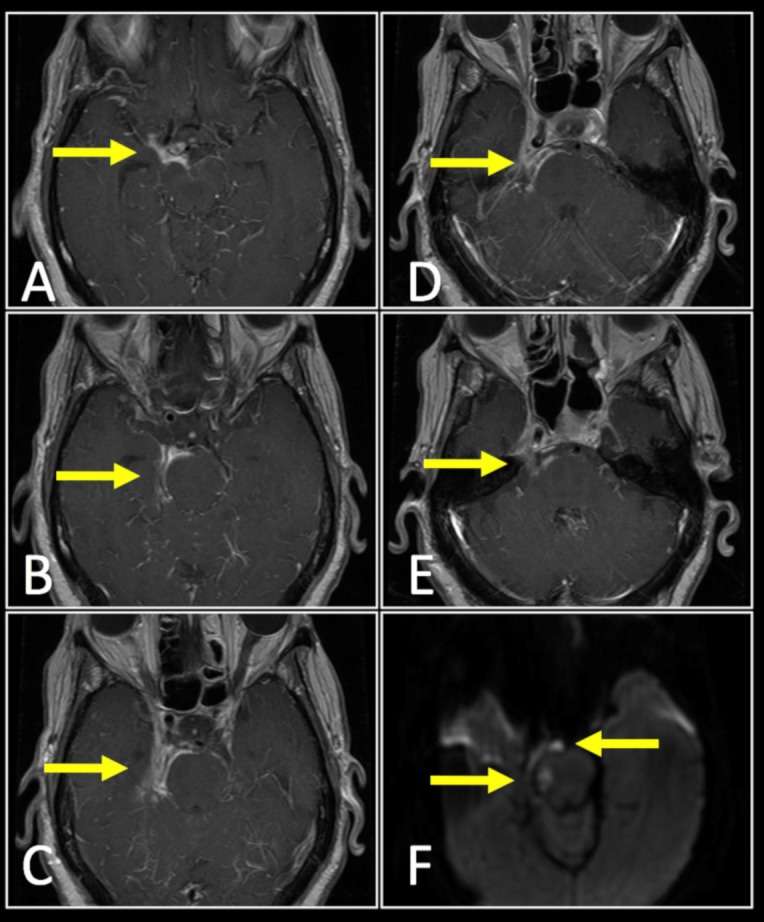
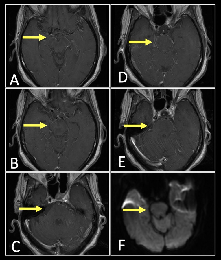
We present a novel treatment with the use of intraventricular antibiotics delivered through a ventriculostomy in a patient who developed septic cavernous sinus thrombosis after sinus surgery. A 65-year-old woman presented with acute on chronic sinusitis. The patient underwent a diagnostic left maxillary antrostomy, ethmoidectomy, sphenoidotomy and sinusotomy. Postoperatively, the patient experienced altered mental status with episodic fever despite treatment with broad-spectrum antimicrobial therapy. MRI of the brain showed extensive meningeal enhancement with the involvement of the right trigeminal and abducens nerve along with thick enhancement along the right pons and midbrain. MR arteriogram revealed a large filling defect within the cavernous sinus. Intraventricular gentamicin was administered via external ventricular drain (ie, ventriculostomy) every 24 hours for 14 days with continued treatment of intravenous ceftriaxone and metronidazole. The patient improved with complete resolution of her cavernous sinus meningitis on repeat brain imaging at 6 months posthospitalisation.
Keywords: drug therapy related to surgery; ear, nose and throat/otolaryngology; infection (neurology); meningitis; neurosurgery.
© BMJ Publishing Group Limited 2019. No commercial re-use. See rights and permissions. Published by BMJ.
Conflict of interest statement
Competing interests: None declared.
Figures
Similar articles
-
Bilateral cavernous sinus thrombosis complicating acute unilateral pansinusitis in a 15-year-old boy.BMJ Case Rep. 2020 Dec 22;13(12):e237758. doi: 10.1136/bcr-2020-237758. BMJ Case Rep. 2020. PMID: 33370987 Free PMC article.
-
Acute sphenoid sinusitis leading to contralateral cavernous sinus thrombosis: a case report.J Laryngol Otol. 2013 Aug;127(8):814-6. doi: 10.1017/S0022215113001527. Epub 2013 Jul 24. J Laryngol Otol. 2013. PMID: 23883649 Review.
-
A review of eight cases of cavernous sinus thrombosis secondary to sphenoid sinusitis, including a12-year-old girl at the present department.Infect Dis (Lond). 2017 Sep;49(9):641-646. doi: 10.1080/23744235.2017.1331465. Epub 2017 May 23. Infect Dis (Lond). 2017. PMID: 28535728 Review.
-
Cavernous sinus thrombosis: successful treatment using functional endonasal sinus surgery.Arch Otolaryngol Head Neck Surg. 1993 Dec;119(12):1368-72. doi: 10.1001/archotol.1993.01880240106014. Arch Otolaryngol Head Neck Surg. 1993. PMID: 17431992
-
Cavernous sinus thrombosis due to ipsilateral sphenoid sinusitis.BMJ Case Rep. 2019 Jan 29;12(1):e227302. doi: 10.1136/bcr-2018-227302. BMJ Case Rep. 2019. PMID: 30700458 Free PMC article.
References
Publication types
MeSH terms
Substances
LinkOut - more resources
Full Text Sources