. 2019 Apr 23;12(578):eaav3810.
doi: 10.1126/scisignal.aav3810.

Tracing the origin and evolution of pseudokinases across the tree of life

Affiliations

Tracing the origin and evolution of pseudokinases across the tree of life

Annie Kwon et al. Sci Signal. .

Abstract

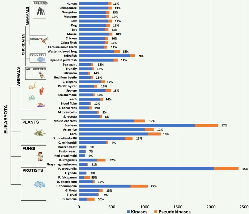
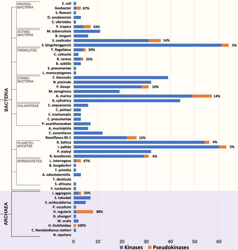
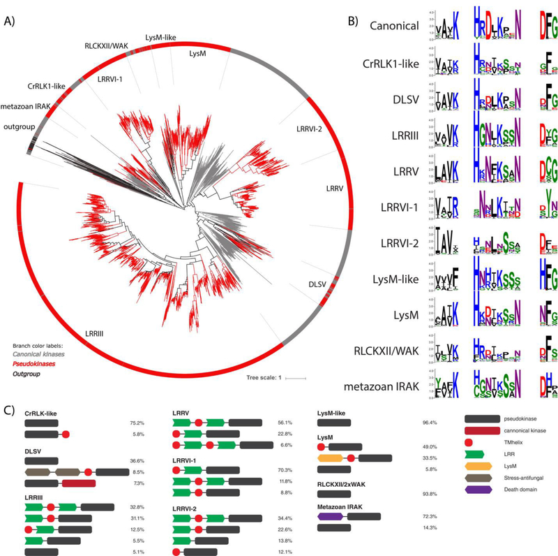
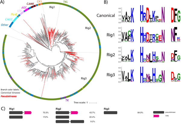
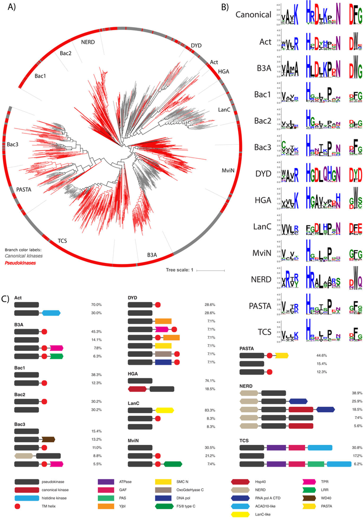
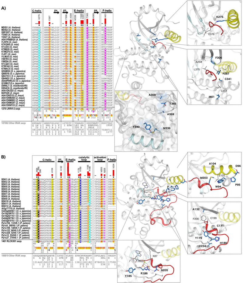
Protein phosphorylation by eukaryotic protein kinases (ePKs) is a fundamental mechanism of cell signaling in all organisms. In model vertebrates, ~10% of ePKs are classified as pseudokinases, which have amino acid changes within the catalytic machinery of the kinase domain that distinguish them from their canonical kinase counterparts. However, pseudokinases still regulate various signaling pathways, usually doing so in the absence of their own catalytic output. To investigate the prevalence, evolutionary relationships, and biological diversity of these pseudoenzymes, we performed a comprehensive analysis of putative pseudokinase sequences in available eukaryotic, bacterial, and archaeal proteomes. We found that pseudokinases are present across all domains of life, and we classified nearly 30,000 eukaryotic, 1500 bacterial, and 20 archaeal pseudokinase sequences into 86 pseudokinase families, including ~30 families that were previously unknown. We uncovered a rich variety of pseudokinases with notable expansions not only in animals but also in plants, fungi, and bacteria, where pseudokinases have previously received cursory attention. These expansions are accompanied by domain shuffling, which suggests roles for pseudokinases in plant innate immunity, plant-fungal interactions, and bacterial signaling. Mechanistically, the ancestral kinase fold has diverged in many distinct ways through the enrichment of unique sequence motifs to generate new families of pseudokinases in which the kinase domain is repurposed for noncanonical nucleotide binding or to stabilize unique, inactive kinase conformations. We further provide a collection of annotated pseudokinase sequences in the Protein Kinase Ontology (ProKinO) as a new mineable resource for the signaling community.


Conflict of interest statement

Competing interests: The authors declare that they have no conflicts of interest with the contents of this article.

Figures

Fig. 1.
Fig. 1.
Kinome and pseudokinome sizes evaluated in 46 eukaryotic species. Blue bars represent the number of kinases detected in the proteome of each species, and orange bars represent the number of pseudokinases. Percentages indicate the fraction of kinases from each proteome that were determined to be pseudokinases. The tree on the left indicates major evolutionary kingdoms and phyla.
Fig. 2.
Fig. 2.
Kinome and pseudokinome sizes evaluated in 51 bacterial and archaeal species. Blue bars represent the number of kinases detected in the proteome of each species, and orange bars represent the number of pseudokinases. Percentages indicate the fraction of kinases from each proteome that were determined to be pseudokinases. The tree on the left indicates major evolutionary kingdoms and phyla.
Fig. 3.
Fig. 3.
A new classification of pseudokinase families. Each colored circle represents a distinct pseudokinase family, which is colored according to the taxonomic group(s) in which it is found. Kinase groups and families from the human kinome classification (18) are depicted by gray circles and labeled in bold font.
Fig. 4.
Fig. 4.
Plant-specific IRAK pseudokinase families. (A) Phylogenetic tree of catalytically active and pseudokinase members of the IRAK family. The 9 plant IRAK pseudokinase families are labeled, and IRAK pseudokinase sequences are shown in red. Canonical IRAK sequences are shown in gray. Outgroup sequences are shown in black. (B) Sequence logos of catalytic motifs for IRAK pseudokinase families. (C) Unique domain structures observed in plant IRAK pseudokinase families. The most common domain structures observed in each family are shown (occurring >5%), with frequencies of each domain structure indicated.
Fig. 5.
Fig. 5.
Rhizophagus irregularis-specific TKL pseudokinase families. (A) Phylogenetic tree of the R. irregularis kinome. Canonical kinase branches are colored in gray and pseudokinases in red. Major kinase groups are labelled using different colors in the outer circle. The 3 major R. irregularis specific pseudokinase families are labelled as Rig1, Rig2 and Rig3. (B) Sequence logos of catalytic motifs for Rig1, Rig2, and Rig3 pseudokinase families. (C) The most common domain structures observed in Rig pseudokinase families are shown (occurring >5%), with frequencies of each domain structure indicated.
Fig. 6.
Fig. 6.
Bacterial pknB pseudokinase families. (A) Phylogenetic tree of pknB canonical kinases and pseudokinases. The 12 pknB-related pseudokinase families are labeled and shown in red branches. Representative canonical pknB kinases are shown in gray. (B) Sequence logos of catalytic motifs in pknB pseudokinase families. (C) Unique domain structures observed in bacterial pseudokinase families. The most common domain structures observed in each family are shown (occurring >5%), with percentages indicating the frequency of each domain structure.
Fig. 7.
Fig. 7.
IRAK pseudokinase-specific features contribute to unique conformations in key catalytic regions. (A) LRRVI-2 pseudokinase family-specific sequence motifs. In the alignment, columns are highlighted where amino acids are highly conserved in LRRVI-2 pseudokinase family sequences and non-conserved and/or biochemically dissimilar in other IRAK sequences. Red bar lengths quantify the degree of divergence between LRRVI-2 and other IRAK sequences. Column-wise amino acid and insertion/deletion frequencies are indicated in integer tenths where a “5” indicates an occurrence of 50–60% in the given (weighted) sequence set. Columns used by the Bayesian partitioning procedure to sort LRRVI-2 sequences from other IRAK sequences are marked with black dots. Kinase secondary structures are annotated above the alignment. In the structure, the glycine-rich loop is colored in light cyan, the C-helix in yellow, and the activation loop in red. Family-specific residues are shown in blue sticks. Residues occurring in canonical catalytic motifs are shown in black lines. Hydrogen bonds are shown in dashed black lines. (B) RLCKXII-1 pseudokinase family-specific sequence motifs.

References

    1. Cohen P, The origins of protein phosphorylation. Nat Cell Biol 4, E127–130 (2002). - PubMed
    1. Kannan N, Taylor SS, Zhai Y, Venter JC, Manning G, Structural and functional diversity of the microbial kinome. PLoS Biol 5, e17 (2007). - PMC - PubMed
    1. Knighton DR et al., Crystal structure of the catalytic subunit of cyclic adenosine monophosphate-dependent protein kinase. Science 253, 407–414 (1991). - PubMed
    1. Knighton DR et al., Structure of a peptide inhibitor bound to the catalytic subunit of cyclic adenosine monophosphate-dependent protein kinase. Science 253, 414–420 (1991). - PubMed
    1. Scheeff ED, Eswaran J, Bunkoczi G, Knapp S, Manning G, Structure of the pseudokinase VRK3 reveals a degraded catalytic site, a highly conserved kinase fold, and a putative regulatory binding site. Structure 17, 128–138 (2009). - PMC - PubMed

Publication types

Substances

LinkOut - more resources