

Proximity biotinylation identifies a set of conformation-specific interactions between Merlin and cell junction proteins
- PMID: 31015291
- PMCID: PMC6631321
- DOI: 10.1126/scisignal.aau8749
Proximity biotinylation identifies a set of conformation-specific interactions between Merlin and cell junction proteins
Abstract
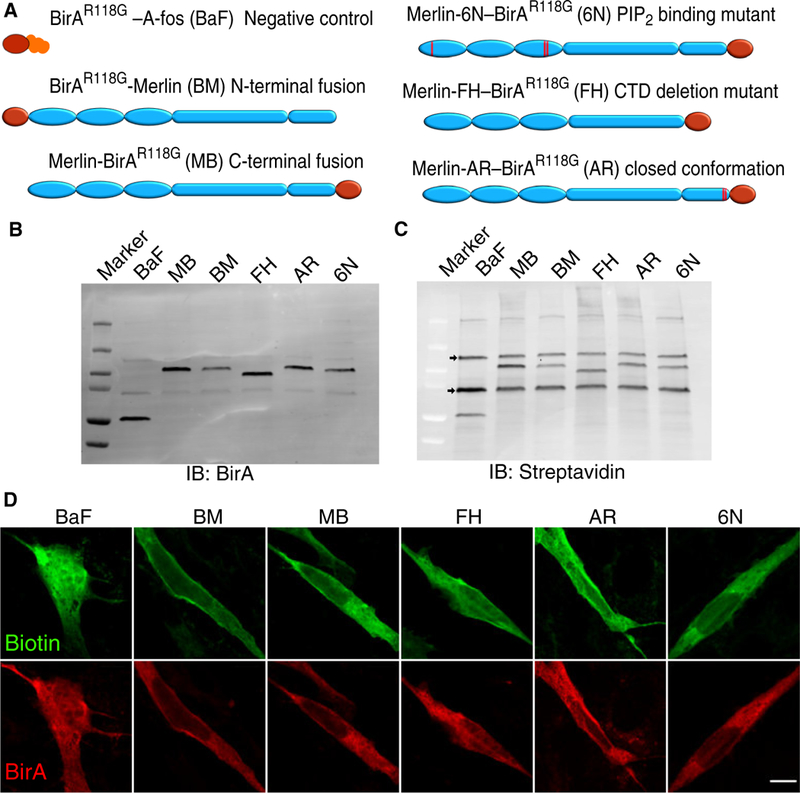
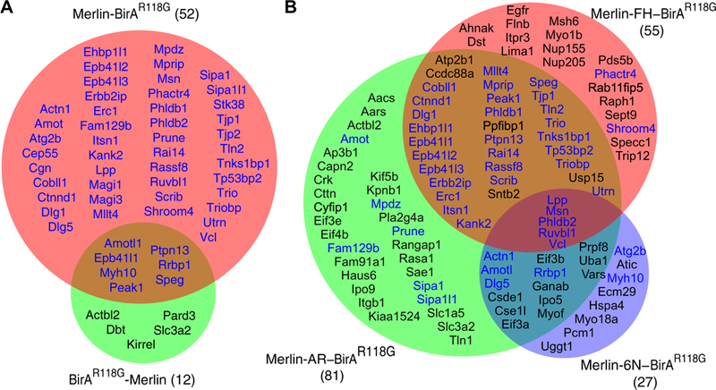
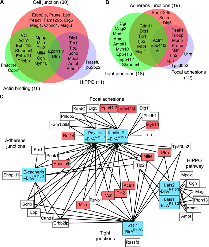
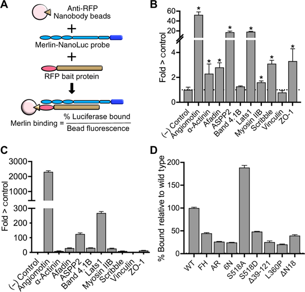
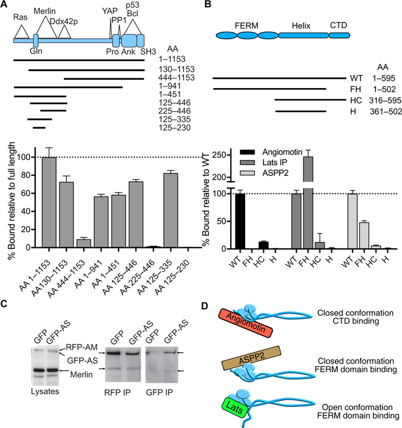
Neurofibromatosis type 2 is an inherited, neoplastic disease associated with schwannomas, meningiomas, and ependymomas and that is caused by inactivation of the tumor suppressor gene NF2 The NF2 gene product, Merlin, has no intrinsic catalytic activity; its tumor suppressor function is mediated through the proteins with which it interacts. We used proximity biotinylation followed by mass spectrometry and direct binding assays to identify proteins that associated with wild-type and various mutant forms of Merlin in immortalized Schwann cells. We defined a set of 52 proteins in close proximity to wild-type Merlin. Most of the Merlin-proximal proteins were components of cell junctional signaling complexes, suggesting that additional potential interaction partners may exist in adherens junctions, tight junctions, and focal adhesions. With mutant forms of Merlin that cannot bind to phosphatidylinositol 4,5-bisphosphate (PIP2) or that constitutively adopt a closed conformation, we confirmed a critical role for PIP2 binding in Merlin function and identified a large cohort of proteins that specifically interacted with Merlin in the closed conformation. Among these proteins, we identified a previously unreported Merlin-binding protein, apoptosis-stimulated p53 protein 2 (ASPP2, also called Tp53bp2), that bound to closed-conformation Merlin predominately through the FERM domain. Our results demonstrate that Merlin is a component of cell junctional mechanosensing complexes and defines a specific set of proteins through which it acts.
Copyright © 2019 The Authors, some rights reserved; exclusive licensee American Association for the Advancement of Science. No claim to original U.S. Government Works.
Figures
Similar articles
-
Inhibition of SIRT2 in merlin/NF2-mutant Schwann cells triggers necrosis.Oncotarget. 2013 Dec;4(12):2354-65. doi: 10.18632/oncotarget.1422. Oncotarget. 2013. PMID: 24259290 Free PMC article.
-
Interaction between two isoforms of the NF2 tumor suppressor protein, merlin, and between merlin and ezrin, suggests modulation of ERM proteins by merlin.J Neurosci Res. 2000 Nov 15;62(4):491-502. doi: 10.1002/1097-4547(20001115)62:4<491::AID-JNR3>3.0.CO;2-D. J Neurosci Res. 2000. PMID: 11070492
-
Merlin tumor suppressor function is regulated by PIP2-mediated dimerization.PLoS One. 2023 Feb 21;18(2):e0281876. doi: 10.1371/journal.pone.0281876. eCollection 2023. PLoS One. 2023. PMID: 36809290 Free PMC article.
-
[Neurofibromatosis type 2 (NF2)].Gan To Kagaku Ryoho. 1997 Sep;24(11):1427-31. Gan To Kagaku Ryoho. 1997. PMID: 9309136 Review. Japanese.
-
A neuronal function of the tumor suppressor protein merlin.Acta Neuropathol Commun. 2014 Jul 12;2:82. doi: 10.1186/s40478-014-0082-1. Acta Neuropathol Commun. 2014. PMID: 25012216 Free PMC article. Review.
Cited by
-
JAM-A signals through the Hippo pathway to regulate intestinal epithelial proliferation.iScience. 2022 Apr 27;25(5):104316. doi: 10.1016/j.isci.2022.104316. eCollection 2022 May 20. iScience. 2022. PMID: 35602956 Free PMC article.
-
Proximity Dependent Biotinylation: Key Enzymes and Adaptation to Proteomics Approaches.Mol Cell Proteomics. 2020 May;19(5):757-773. doi: 10.1074/mcp.R120.001941. Epub 2020 Mar 3. Mol Cell Proteomics. 2020. PMID: 32127388 Free PMC article. Review.
-
MerlinS13 phosphorylation regulates meningioma Wnt signaling and magnetic resonance imaging features.Nat Commun. 2024 Sep 9;15(1):7873. doi: 10.1038/s41467-024-52284-8. Nat Commun. 2024. PMID: 39251601 Free PMC article.
-
HPV18 E7 inhibits LATS1 kinase and activates YAP1 by degrading PTPN14.bioRxiv [Preprint]. 2024 Jun 19:2024.03.07.583953. doi: 10.1101/2024.03.07.583953. bioRxiv. 2024. Update in: mBio. 2024 Oct 16;15(10):e0181124. doi: 10.1128/mbio.01811-24. PMID: 38496413 Free PMC article. Updated. Preprint.
-
Elucidation of Short Linear Motif-Based Interactions of the FERM Domains of Ezrin, Radixin, Moesin, and Merlin.Biochemistry. 2023 Jun 6;62(11):1594-1607. doi: 10.1021/acs.biochem.3c00096. Epub 2023 May 24. Biochemistry. 2023. PMID: 37224425 Free PMC article.
References
-
- Rouleau GA, Merel P, Lutchman M, Sanson M, Zucman J, Marineau C, Hoang-Xuan K, Demczuk S, Desmaze C, Plougastel B, Pulst SM, Lenoir G, Bijlsma E, Fashold R, Dumanski J, de Jong P, Parry D, Eldrige R, Aurias A, Delattre O, Thomas G, Alteration in a new gene encoding a putative membrane-organizing protein causes neuro-fibromatosis type 2. Nature 363, 515–521 (1993). - PubMed
-
- MacCollin M, Mohney T, Trofatter J, Wertelecki W, Ramesh V, Gusella J, DNA diagnosis of neurofibromatosis 2. Altered coding sequence of the merlin tumor suppressor in an extended pedigree. JAMA 270, 2316–2320 (1993). - PubMed
-
- Ammoun S, Hanemann CO, Emerging therapeutic targets in schwannomas and other merlin-deficient tumors. Nat. Rev. Neurol. 7, 392–399 (2011). - PubMed
Publication types
MeSH terms
Substances
Grants and funding
LinkOut - more resources
Full Text Sources
Molecular Biology Databases
Research Materials
Miscellaneous
