. 2019 Apr 23;10(2):e00297-19.
doi: 10.1128/mBio.00297-19.

Quantitative Analyses Reveal Novel Roles for N- Glycosylation in a Major Enteric Bacterial Pathogen

Affiliations

Quantitative Analyses Reveal Novel Roles for N- Glycosylation in a Major Enteric Bacterial Pathogen

Sherif Abouelhadid et al. mBio. .

Abstract

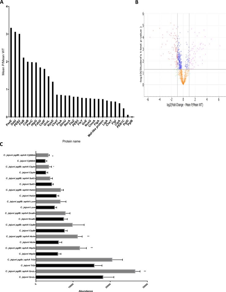
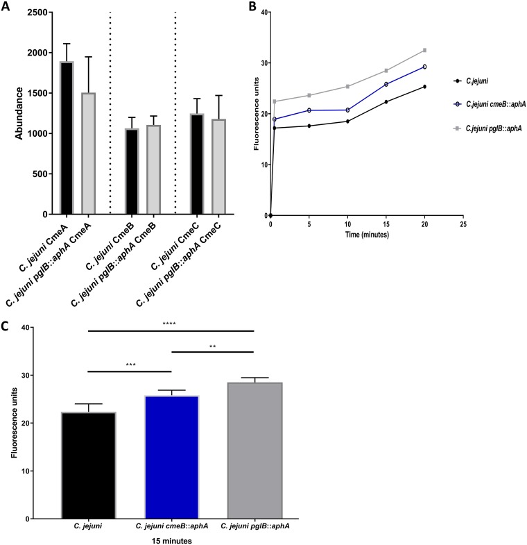
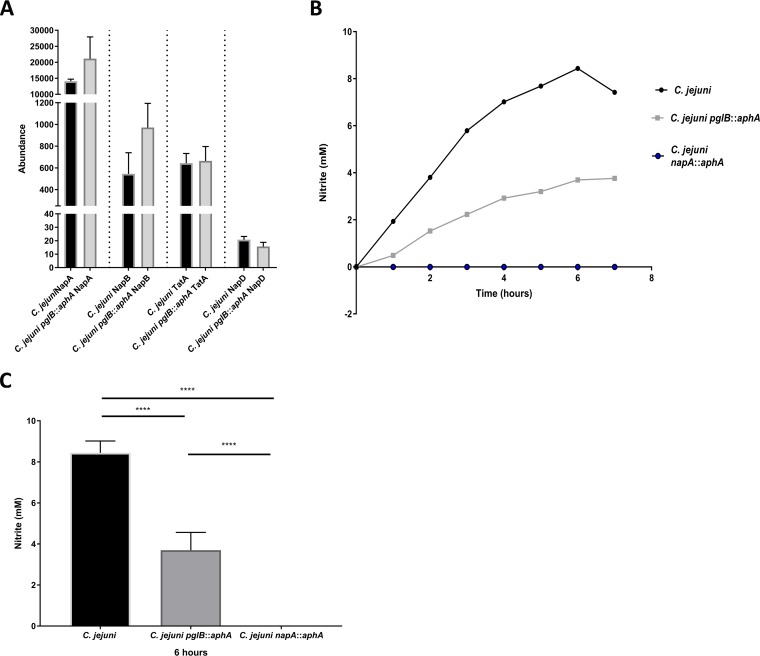
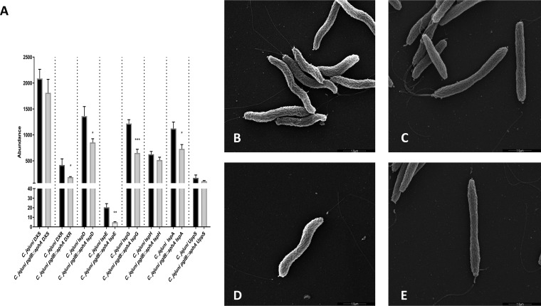
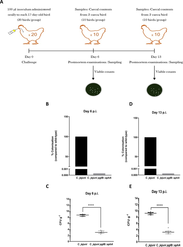
In eukaryotes, glycosylation plays a role in proteome stability, protein quality control, and modulating protein function; however, similar studies in bacteria are lacking. Here, we investigate the roles of general protein glycosylation systems in bacteria using the enteropathogen Campylobacter jejuni as a well-defined example. By using a quantitative proteomic strategy, we were able to monitor changes in the C. jejuni proteome when glycosylation is disrupted. We demonstrate that in C. jejuni, N-glycosylation is essential to maintain proteome stability and protein quality control. These findings guided us to investigate the role of N-glycosylation in modulating bacterial cellular activities. In glycosylation-deficient C. jejuni, the multidrug efflux pump and electron transport pathways were significantly impaired. We demonstrate that in vivo, fully glycosylation-deficient C. jejuni bacteria were unable to colonize its natural avian host. These results provide the first evidence of a link between proteome stability and complex functions via a bacterial general glycosylation system.IMPORTANCE Advances in genomics and mass spectrometry have revealed several types of glycosylation systems in bacteria. However, why bacterial proteins are modified remains poorly defined. Here, we investigated the role of general N-linked glycosylation in a major food poisoning bacterium, Campylobacter jejuni The aim of this study is to delineate the direct and indirect effects caused by disrupting this posttranslational modification. To achieve this, we employed a quantitative proteomic strategy to monitor alterations in the C. jejuni proteome. Our quantitative proteomic results linked general protein N-glycosylation to maintaining proteome stability. Functional analyses revealed novel roles for bacterial N-glycosylation in modulating multidrug efflux pump, enhancing nitrate reduction activity, and promoting host-microbe interaction. This work provides insights on the importance of general glycosylation in proteins in maintaining bacterial physiology, thus expanding our knowledge of the emergence of posttranslational modification in bacteria.

Keywords: glycosylation; host-microbe interaction; microbial physiology; pathogenesis; proteomics.


Figures

FIG 1
FIG 1
Quantitative comparison of wild-type C. jejuni and C. jejuni pglB::aphA proteomes. (A) Selected proteins that are statistically differentially expressed in wild-type C. jejuni and C. jejuni pglB::aphA proteomes. Proteins were analyzed by Perseus and presented according to their fold change with 95% CI. (B) Volcano plot analysis of significantly abundant proteins. The −log10 (Student’s t test) is plotted against log2 mean fold change of C. jejuni pglB::aphA,P C. jejuni, WT. The nonaxial horizontal line denotes P = 0.05, while the nonaxial vertical lines denote 0.5- and 2-fold changes, respectively. The volcano plot was generated using PANDA-view (12). (C) Differential expression of chaperones and proteases in wild-type C. jejuni and C. jejuni pglB::aphA. Data are from three biological replicates. The error bars represent standard deviations. Values that are significantly different by Student’s t test are indicated by asterisks as follows: *, P < 0.05; **, P < 0.01.
FIG 2
FIG 2
Differential expression and functional analysis of CmeABC in C. jejuni. (A) Differential expression of CmeABC in wild-type C. jejuni and C. jejuni pglB::aphA. Data are from three biological replicates, and error bars represent standard deviations. Data were analyzed by Student’s t test. (B) Ethidium bromide accumulation test in C. jejuni strains. Thirty milliliters of brucella broth was separately inoculated with overnight cultures of C. jejuni (black circles), C. jejuni cmeB::aphA (open circles), and C. jejuni pglB::aphA (gray squares) to an OD600 of 0.1. Cells were grown until an OD600 of 0.4 to 0.5 was reached, spun down, washed, and resuspended to an OD600 of 0.2 in 10 mM sodium phosphate buffer (pH 7). Cells were then incubated in a VAIN for 15 min at 37°C, and then ethidium bromide was added to a final concentration of 0.2 mg/ml. Fluorescence was read at excitation and emission for 20 min at 37°C. (B) Ethidium bromide accumulation in C. jejuni strains throughout 20 min. (C) Ethidium bromide accumulation in C. jejuni strains at 15 min. The data are means for three biological replicates, two technical replicates each, and the error bars represent standard deviations. Significance was calculated using one-way ANOVA test with multiple comparison and indicated by asterisks as follows: **, P < 0.01; ***, P < 0.001; ****, P < 0.0001.
FIG 3
FIG 3
Differential expression and functional analysis of NapAB complex in C. jejuni. (A) Differential expression of NapAB, TatA, and NapD in C. jejuni and C. jejuni pglB::aphA. Data are from three biological replicates, and error bars represent standard deviations. Data were analyzed by Student’s t test. (B) Nitrate reduction in C. jejuni strains. Ninety milliliters of brucella broth supplemented with 200 mM nitrate was inoculated separately with wild-type C. jejuni (black circles), C. jejuni pglB::aphA (gray squares), and C. jejuni napA::aphA (blue circles) to an OD600 of 0.1 from an overnight culture. Cultures were incubated statically in a VAIN at 37°C. Samples were withdrawn every hour, C. jejuni cells were removed by centrifugation, and nitrate reduction was measured straight from the supernatant against nitrite standards. (B) Nitrite accumulation in the supernatant over 7 h. (C) Nitrite accumulation in the supernatant at 6 h. The data represents the mean of three biological replicates and two technical replicates, and error bars represent standard deviations. Significance was calculated using one-way ANOVA test with multiple comparison. ****, P < 0.0001.
FIG 4
FIG 4
Abundance of undecaprenyl phosphate biosynthetic enzymes and cell morphology in C. jejuni strains. (A) Differential expression of MEP pathway enzymes and UppS. Data are from three biological replicates, and error bars represent standard deviations. Data were analyzed by Student’s t test. *, P < 0.05; **, P < 0.01; ***, P < 0.001. (B to E) Scanning electron micrographs of wild-type C. jejuni 11168H (B) and C. jejuni 11168H pglB::aphA (C) and of a single bacterial cell of wild-type C. jejuni 11168H (D) and a single bacterial cell of C. jejuni 11168H pglB::aphA (E). The bars represent 1.0 μm.
FIG 5
FIG 5
Colonization of C. jejuni and C. jejuni pglB::aphA in 17-day-old White Leghorn chickens. (A) Schematic diagram of the chicken colonization experiment. Seventeen-day-old chicken were inoculated with 100 μl of 106 CFU of wild-type C. jejuni or C. jejuni pglB::aphA. Chickens were then sacrificed at day 6 and day 13, and postmortem examination (PM) was carried out by directly enumerating C. jejuni on CCDA plates. (B) CFU counts of C. jejuni strains on CCDA plates on day 6 postinoculation. (C) Percentage of C. jejuni strains colonization on day 6 postinoculation. (D) CFU counts of C. jejuni strains on CCDA plates on day 13 postinoculation. (E) Percentage of C. jejuni strain colonization on day 13 postinoculation. Statistical significance was calculated using Mann-Whitney test. ****, P < 0.0001.

References

    1. Varki A. 2017. Biological roles of glycans. Glycobiology 27:3–49. doi: 10.1093/glycob/cww086. - DOI - PMC - PubMed
    1. Linton D, Allan E, Karlyshev AV, Cronshaw AD, Wren BW. 2002. Identification of N-acetylgalactosamine-containing glycoproteins PEB3 and CgpA in Campylobacter jejuni. Mol Microbiol 43:497–508. doi: 10.1046/j.1365-2958.2002.02762.x. - DOI - PubMed
    1. Vik A, Aas FE, Anonsen JH, Bilsborough S, Schneider A, Egge-Jacobsen W, Koomey M. 2009. Broad spectrum O-linked protein glycosylation in the human pathogen Neisseria gonorrhoeae. Proc Natl Acad Sci U S A 106:4447–4452. doi: 10.1073/pnas.0809504106. - DOI - PMC - PubMed
    1. Lithgow KV, Scott NE, Iwashkiw JA, Thomson ELS, Foster LJ, Feldman MF, Dennis JJ. 2014. A general protein O-glycosylation system within the Burkholderia cepacia complex is involved in motility and virulence. Mol Microbiol 92:116–137. doi: 10.1111/mmi.12540. - DOI - PubMed
    1. Fletcher CM, Coyne MJ, Villa OF, Chatzidaki-Livanis M, Comstock LE. 2009. A general O-glycosylation system important to the physiology of a major human intestinal symbiont. Cell 137:321–331. doi: 10.1016/j.cell.2009.02.041. - DOI - PMC - PubMed

Publication types